Просмотр новости
Найдите то, что Вас интересует
 Спутники
 РН
РН SpaceX
 Роскосмос
 NASA
NASA
СпутникиРНSpaceXРоскосмосNASAProtein–protein interactions play a central role in cellular signaling, immune recognition, and therapeutic development, yet they are often characterized by weak affinities, transient binding, and pronounced conformational flexibility. These features present significant challenges for conventional structural biology techniques. Biophysical approaches, particularly nuclear magnetic resonance (NMR) spectroscopy, and isothermal titration calorimetry (ITC) have emerged as powerful tools for elucidating protein-protein interactions in solution. NMR enables residue-specific mapping of interaction interfaces and characterization of conformational dynamics across multiple timescales, making it particularly suited for studying weak and transient interactions. Although performed in solution, NMR experiments typically require elevated protein concentrations and therefore provide a solution-state rather than strictly physiological view of interactions. In contrast, ITC provides a direct and label-free measurement of binding thermodynamics, yielding quantitative parameters such as affinity, stoichiometry, and entropic contributions to binding. This review highlights the principles, applications, strengths, and limitations of NMR and ITC in protein-protein interaction research, emphasizing how their combined use enables an integrated understanding of structure, dynamics, and energetics. Representative examples from the literature are discussed, illustrating the unique ability of NMR and ITC to capture structural and dynamic features of peptide recognition that are critical for understanding biological function and guiding peptide-based therapeutic design.

Ammous-Boukhris N, Gargouri A and Mokdad-Gargouri R. Understanding Protein–Protein Interactions in Biology: Applications of NMR Spectroscopy and Isothermal Titration Calorimetry [version 2; peer review: 1 not approved]. F1000Research 2026, 15:66 (https://doi.org/10.12688/f1000research.176780.2)
Review
Revised
[version 2; peer review: 1 not approved]
https://orcid.org/0000-0002-5744-0328
1, Ali Gargouri1, Raja Mokdad-Gargourihttps://orcid.org/0000-0003-1319-0061
1https://orcid.org/0000-0002-5744-0328
1, Ali Gargouri1, Raja Mokdad-Gargourihttps://orcid.org/0000-0003-1319-0061
11 Center of Biotechnology of Sfax, Laboratory of Eukaryotes Molecular Biotechnology, University of Sfax, Sfax, Tunisia
Nihel Ammous-Boukhris
Roles: Conceptualization, Writing – Original Draft Preparation, Writing – Review & Editing
Ali Gargouri
Roles: Conceptualization, Writing – Review & Editing
Raja Mokdad-Gargouri
Roles: Conceptualization, Writing – Original Draft Preparation, Writing – Review & Editing
OPEN PEER REVIEW
REVIEWER STATUS
Protein–protein interactions play a central role in cellular signaling, immune recognition, and therapeutic development, yet they are often characterized by weak affinities, transient binding, and pronounced conformational flexibility. These features present significant challenges for conventional structural biology techniques. Biophysical approaches, particularly nuclear magnetic resonance (NMR) spectroscopy, and isothermal titration calorimetry (ITC) have emerged as powerful tools for elucidating protein-protein interactions in solution. NMR enables residue-specific mapping of interaction interfaces and characterization of conformational dynamics across multiple timescales, making it particularly suited for studying weak and transient interactions. Although performed in solution, NMR experiments typically require elevated protein concentrations and therefore provide a solution-state rather than strictly physiological view of interactions. In contrast, ITC provides a direct and label-free measurement of binding thermodynamics, yielding quantitative parameters such as affinity, stoichiometry, and entropic contributions to binding. This review highlights the principles, applications, strengths, and limitations of NMR and ITC in protein-protein interaction research, emphasizing how their combined use enables an integrated understanding of structure, dynamics, and energetics. Representative examples from the literature are discussed, illustrating the unique ability of NMR and ITC to capture structural and dynamic features of peptide recognition that are critical for understanding biological function and guiding peptide-based therapeutic design.
Nuclear Magnetic Resonance, Isothermal Titration Calorimetry, Peptide, Interaction, Biophysical approaches
Corresponding author: Raja Mokdad-Gargouri Competing interests: No competing interests were disclosed.
Grant information: This research is funded by the Tunisian Ministry of Higher Education and Scientific Research (Grant: LR19/CBS02).
The funders had no role in study design, data collection and analysis, decision to publish, or preparation of the manuscript.
Copyright: © 2026 Ammous-Boukhris N et al. This is an open access article distributed under the terms of the Creative Commons Attribution License, which permits unrestricted use, distribution, and reproduction in any medium, provided the original work is properly cited. How to cite: Ammous-Boukhris N, Gargouri A and Mokdad-Gargouri R. Understanding Protein–Protein Interactions in Biology: Applications of NMR Spectroscopy and Isothermal Titration Calorimetry [version 2; peer review: 1 not approved]. F1000Research 2026, 15:66 (https://doi.org/10.12688/f1000research.176780.2) First published: 16 Jan 2026, 15:66 (https://doi.org/10.12688/f1000research.176780.1) Latest published: 27 Jul 2026, 15:66 (https://doi.org/10.12688/f1000research.176780.2)
The revised version of the manuscript incorporates the corrections suggested by the reviewer.
Major amendments from version 1 include the substantial expansion of the methodological sections dedicated to NMR spectroscopy and ITC, with clearer explanations of CSP, STD-NMR, relaxation measurements, exchange regimes, and thermodynamic analysis. Numerous recent and representative references were integrated to strengthen the scientific depth of the review and provide detailed methodological illustrations. The application section was extensively reorganised and enriched with mechanistic examples combining NMR and ITC approaches. In addition, figures, legends, and the graphical abstract were added to improve clarity, readability, and overall presentation of the manuscript.
See the authors' detailed response to the review by Barbara Zambelli
Protein–protein interactions (PPIs) are central to all biological processes, including signal transduction, enzymatic regulation, transcriptional control, and cellular architecture.1 Rather than acting as isolated entities, proteins function within complex and dynamic interaction networks, where the specificity, strength, and regulation of PPIs determine biological outcomes.2,3 Understanding these interactions at the molecular level is therefore essential for elucidating biological mechanisms and for developing therapeutic strategies targeting dysregulated protein interactions.
Traditional approaches to studying PPIs, such as yeast two-hybrid assays4 or co-immunoprecipitation,5 are powerful for identifying interaction partners but provide limited quantitative or mechanistic information. In contrast, biophysical techniques offer direct insight into the molecular basis of PPIs by characterizing binding affinities, thermodynamic driving forces, structural interfaces, and conformational dynamics.6,7 Among these techniques, nuclear magnetic resonance (NMR) spectroscopy and isothermal titration calorimetry (ITC) have emerged as particularly valuable tools.
NMR spectroscopy enables the study of PPIs at atomic resolution in solution, allowing researchers to map interaction interfaces, detect conformational changes, and characterize weak or transient interactions that are often inaccessible to crystallographic methods. Importantly, NMR does not require immobilization or crystallization, thereby preserving the native conformational ensemble, although it typically operates at protein concentrations higher than physiological levels.8 Complementing this structural and dynamic information, ITC provides a direct and label-free measurement of the thermodynamic parameters governing protein binding, including binding affinity, enthalpy, entropy, and stoichiometry.9,10
This review focuses on the application of NMR spectroscopy and ITC to the study of protein-protein interactions in biological research. Particular emphasis is placed on how these techniques are applied in practice, their methodological limitations, and how their integration provides mechanistic insight into molecular recognition.
NMR spectroscopy exploits the magnetic properties of atomic nuclei to obtain detailed information about the structure and environment of biomolecules in solution. In biomolecular NMR, titration experiments enables residue-specific assignment and atomic-resolution analysis of protein interactions.11 Unlike many structural techniques that provide static snapshots, NMR is inherently sensitive to molecular motions across a wide range of timescales.12 This allows direct characterization of conformational exchange processes, which are often central to binding mechanisms such as induced fit or conformational selection.
Chemical shift perturbation (CSP) experiments remain a cornerstone of NMR PPI analysis. In these experiments, changes in NMR resonance frequencies are monitored as one protein is titrated with its binding partner.13 Residues located at or near the binding interface experience changes in their local chemical environment, resulting in measurable chemical shift changes.12,14 By mapping these perturbations onto the protein’s three-dimensional structure or sequence, researchers can identify interaction surfaces with residue-level resolution. This approach is particularly valuable for guiding mutagenesis experiments, validating interaction models, and distinguishing direct binding interfaces from allosteric effects.
Importantly, the interpretation of CSP data depends strongly on the exchange regime. In the fast exchange regime, peaks shift continuously, allowing quantitative estimation of the dissociation constant (Kd). In the intermediate exchange regime, peaks broaden, which typically indicates micromolar to millimolar affinity and ongoing conformational exchange. In the slow exchange regime, separate peaks are observed for the free and bound states, reflecting the presence of distinct populations ( Figure 1).8
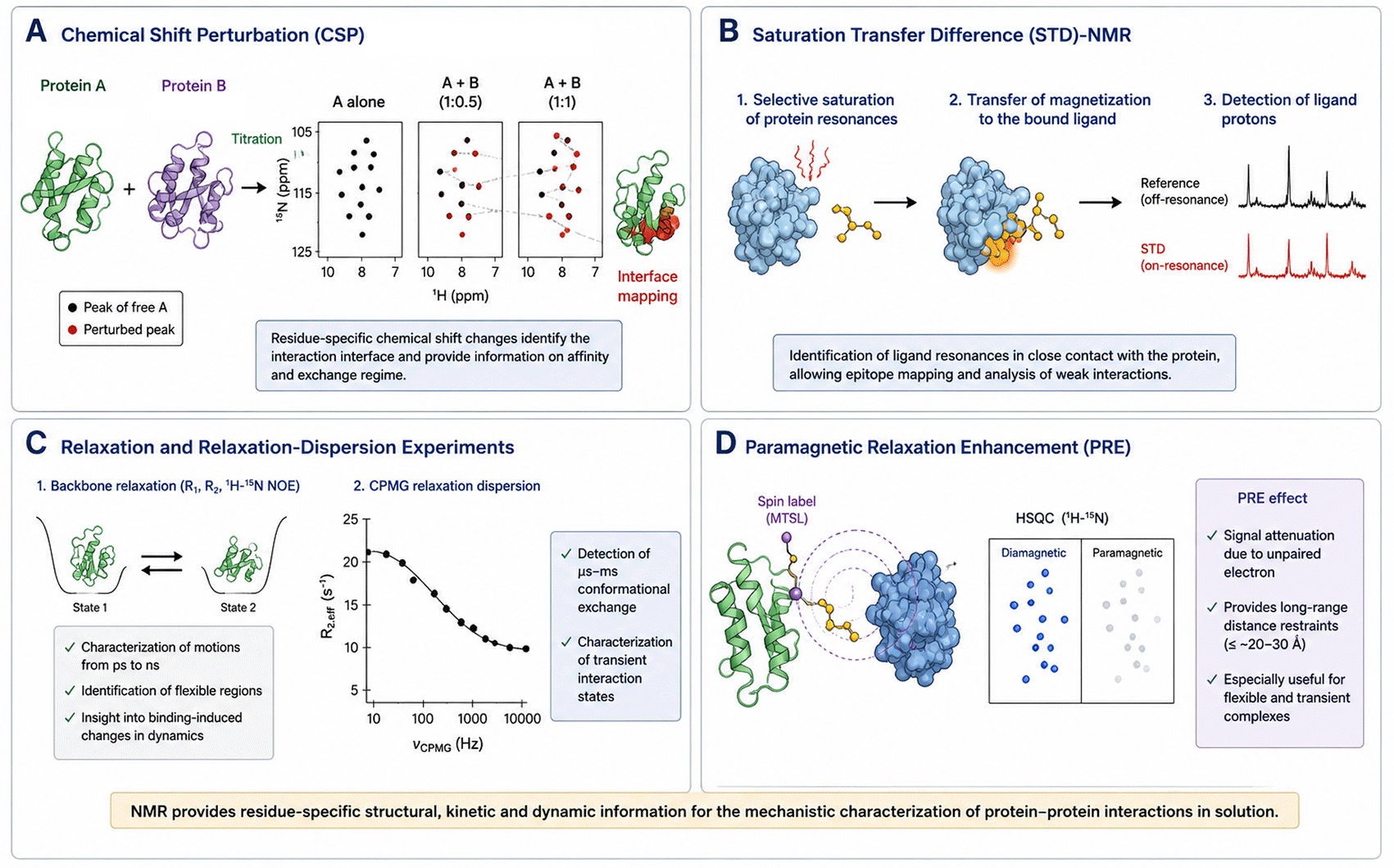
Schematic representation of the main NMR methodologies used to characterize protein–protein interactions (PPIs). (A) Chemical shift perturbation (CSP) analysis using titration experiments, where residue-specific peak shifts identify interaction interfaces and provide information on binding affinity and exchange regime. (B) Saturation Transfer Difference (STD)-NMR workflow illustrating selective saturation of the protein followed by magnetization transfer to the bound ligand, enabling epitope mapping of interacting residues. (C) Relaxation and relaxation-dispersion experiments used to investigate protein dynamics, conformational exchange, and transient interaction states. (D) Integration of NMR-derived restraints with computational modeling to generate structural ensembles of dynamic protein complexes. Together, these approaches provide complementary structural, kinetic, and dynamic information for the mechanistic characterization of PPIs.
Furthermore, integrative strategies combining NMR-derived restraints with computational docking approaches are increasingly used to reconstruct the quaternary structure of protein complexes. In such workflows, CSP and distance restraints define interaction surfaces, while modeling algorithms generate structural ensembles consistent with experimental data.
Recent applications, such as NMR titration studies of AcpP-mediated protein–protein interactions in the E. coli FAS-II system, demonstrate how CSP combined with line-shape analysis can provide both binding affinities and mechanistic insight into transient enzymatic complexes. In these systems, interface mapping was complemented by kinetic information derived from exchange behavior, highlighting how NMR moves beyond simple localization toward quantitative mechanistic interpretation.15–17
Ligand-observed techniques such as Saturation Transfer Difference (STD) NMR are particularly valuable for weak interactions and screening applications.17
STD-NMR works by selectively saturating protein signals and transferring this saturation to bound ligands. Only ligand protons in close contact with the protein receive this signal, allowing direct identification of binding epitopes. This makes it especially suitable for low-affinity interactions (μM–mM range) where classical methods struggle.18,19
Recent methodological advances have significantly expanded the efficiency of STD-NMR:
Imaging STD-NMR enables simultaneous evaluation of binding affinity and specificity within a single experiment, allowing spatially resolved analysis of ligand interactions and reducing experimental complexity.15
The reduced data set STD (rd-STD) NMR approach enables faster affinity determination using fewer spectra, making it suitable for high-throughput screening.20
Importantly, STD-NMR is not suited for interactions between two large folded proteins, but is highly effective for protein–peptide and protein–small molecule systems.20
Many biologically relevant PPIs are characterized by low affinities and short lifetimes, such as those involved in signaling cascades or regulatory processes. These interactions are often difficult to capture using crystallography or cryo-electron microscopy. NMR, however, is well suited to detect and characterize such interactions because it operates at high protein concentrations (typically 50–500 μM), enabling detection of weak interactions, and it captures exchange processes between free and bound states.15 However, these conditions may also promote detection of non-specific interactions, which must be carefully controlled.
For example, NMR studies of the transcription factor MntR from Mycobacterium tuberculosis showed how metal binding induces conformational changes that regulate protein interactions, highlighting the sensitivity of NMR to subtle structural rearrangements.21 Techniques such as line broadening analysis, relaxation measurements, and exchange spectroscopy allow quantitative analysis of binding kinetics and transient interaction states, including encounter complexes.15
Beyond mapping interfaces, NMR provides unique insights into how PPIs influence conformational dynamics.16 These dynamic effects are often central to biological function.17 Relaxation measurements (R1, R2, NOE) and relaxation dispersion experiments allow characterization of motions spanning picoseconds to milliseconds, which are often directly linked to biological function.
In dynamic PPIs, NMR can distinguish between conformational selection and induced fit mechanisms, as well as identify allosteric communication pathways across protein structures. This is particularly important for Intrinsically disordered proteins (IDPs), Multi-domain regulatory proteins, and Transient signaling complexes.22 For instance, the interaction between the transactivation domain of N-Myc and its partner TFIIIC5 was shown to involve disorder-to-order transitions upon binding, illustrating a coupled folding–binding mechanism typical of transcriptional regulators.23 Similarly, phosphorylation-dependent modulation of IDR interactions has been characterized by NMR, as demonstrated for the oncogenic protein NDRG1, where phosphorylation disrupts membrane association by altering conformational dynamics and electrostatic interactions.24
NMR offers several key advantages for the study of PPIs, including atomic scale resolution, sensitivity to dynamics, and the ability to study proteins in solution under physiologically relevant conditions. It is particularly powerful for analyzing weak, transient, and disordered interactions that are difficult to access using other techniques.
However, NMR also has limitations. The size of protein complexes that can be studied is constrained by spectral complexity and sensitivity, although advances such as transverse relaxation-optimized spectroscopy (TROSY) and higher magnetic field strengths have significantly extended these limits.25 In addition, NMR experiments often require high sample concentration, which can limit throughput.26
Isothermal titration calorimetry (ITC) is a label-free biophysical technique that directly measures the heat exchanged during molecular binding events.26 In ITC experiments, one protein is titrated into a solution containing its binding partner, and the resulting heat changes are recorded as a function of molar ratio. From a single experiment, ITC provides a complete thermodynamic profile of the interaction, including the binding affinity (Kd), enthalpy change (ΔH), entropy change (ΔS), and binding stoichiometry (n).26
Interestingly, the practical interpretation of ITC data requires careful consideration of experimental design and binding models. The shape of the thermogram and resulting binding isotherm depends not only on affinity but also on concentration regimes (c-value), injection strategy, and heat of dilution corrections. Accurate ITC analysis requires careful experimental design, including buffer matching, concentration optimization (c-value), and appropriate binding models for data fitting.
In the context of PPIs, ITC is particularly valuable because it measures binding energetics directly, without relying on fluorescent labels, surface immobilization, or indirect readouts. This directness makes ITC a gold-standard method for quantifying PPIs and for validating interactions identified by other biochemical or biophysical techniques.27
A key strength of ITC in PPI research is its ability to accurately determine binding affinities over a broad range, from weak micromolar interactions to tight nanomolar complexes, provided that appropriate experimental conditions are used.27 The determination of binding stoichiometry is especially important for protein complexes that may form higher-order assemblies or oligomeric states, as ITC can distinguish between different binding models. This quantitative information is critical for understanding biological function, as the strength and stoichiometry of protein interactions often dictate signaling thresholds, complex formation, and regulatory mechanisms within the cell.28
Beyond affinity measurements, ITC uniquely provides insight into the thermodynamic forces that drive PPIs. By separating the contributions of enthalpy (ΔH) and entropy (ΔS), ITC allows researchers to understand the molecular origin of binding. Enthalpy-driven interactions are often associated with the formation of specific non-covalent contacts such as hydrogen bonds and electrostatic interactions, whereas entropy-driven interactions may reflect hydrophobic effects, solvent release, or conformational changes.29
For example, ITC studies of trypsin and its peptidic inhibitors showed that binding is mainly enthalpy-driven, due to strong intermolecular interactions, while entropy penalties arise from reduced flexibility upon complex formation.30 A second example, ITC analysis of flavan-3-ol interactions with α-amylase revealed how polyphenolic compounds modulate enzyme activity through specific binding modes, with thermodynamic signatures correlating with inhibitory effects on starch hydrolysis.31
Such analyses illustrate how ITC can go beyond affinity determination to reveal the molecular origin of binding specificity and strength, which is particularly important when comparing related ligands or engineered variants.
ITC is widely used in mutational analyses of protein-protein interfaces, where point mutations are introduced to assess their impact on binding energetics.32,33,35,36 By comparing thermodynamic parameters between wild-type and mutant proteins, researchers can distinguish between residues that contribute directly to binding affinity and those that influence interaction stability indirectly. For example, studies on engineered binding proteins (such as monobodies interacting with adenylate kinase) showed how specific mutations alter binding affinity and specificity, directly linking structural changes to energetic effects.34 Additionally, ITC is frequently employed in comparative studies of homologous proteins or interaction partners, enabling the identification of evolutionary or functional differences in binding mechanisms.35
While ITC is widely used for soluble proteins, its application to membrane-associated systems presents additional challenges.
Membrane proteins require detergents or lipid mimetics for solubility, and heat signals may include contributions from lipid reorganization, making data interpretation more complex. For example, ITC studies of the FNR–cytochrome b6f complex demonstrated that careful experimental design allows reliable measurement of binding energetics even in heterogeneous membrane environments.36
Despite these challenges, ITC remains applicable and highly informative for membrane-associated PPIs when conditions are properly controlled.
The principal advantage of ITC is its ability to provide a complete thermodynamic description of PPIs in a single experiment, using native proteins and solution conditions that closely mimic the cellular environment. This makes ITC an essential complement to structural techniques such as NMR and crystallography. However, ITC also has limitations. The technique typically requires relatively large amounts of highly purified protein, which can be challenging for unstable or low-yield systems.9 In addition, ITC provides limited structural information and is less sensitive to very weak interactions without careful experimental optimization.9 As a result, ITC is most powerful when used in combination with structural and spectroscopic methods.
The combination of NMR experiments and ITC measurements provides a powerful framework for understanding PPIs at multiple levels ( Figure 2).
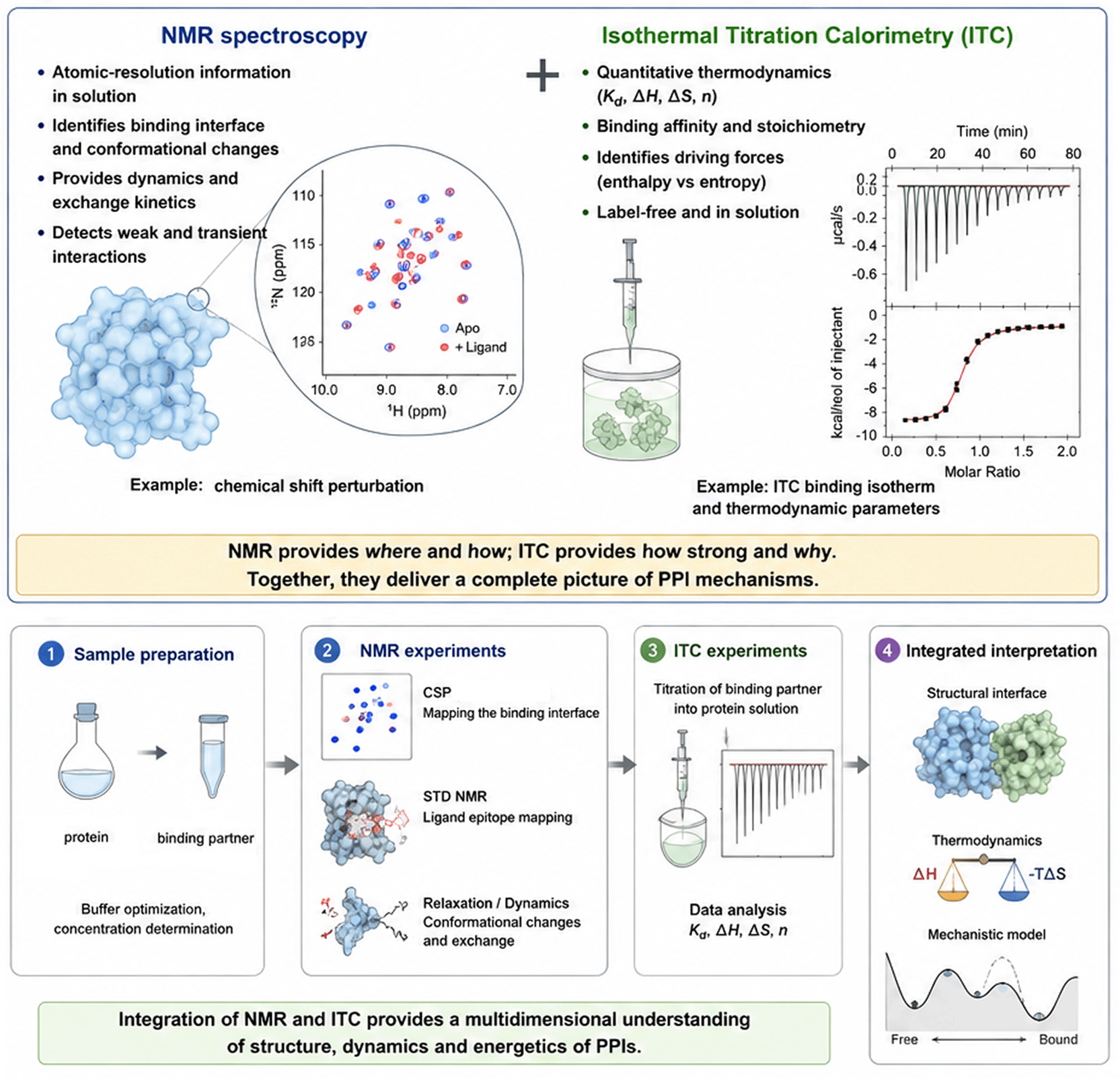
Integrated biophysical strategy combining NMR spectroscopy and isothermal titration calorimetry (ITC) to characterize protein–protein interactions. The workflow begins with sample preparation, followed by NMR experiments including CSP mapping, STD-NMR, and relaxation measurements to identify binding interfaces, characterize dynamics, and detect transient interactions. Subsequently, ITC experiments quantify the thermodynamic parameters of binding, including dissociation constant (Kd), binding stoichiometry (n), enthalpy (ΔH), and entropy (ΔS). The integration of structural, dynamic, kinetic, and thermodynamic data enables the construction of comprehensive mechanistic models of molecular recognition and interaction specificity.
To illustrate how NMR spectroscopy and isothermal titration calorimetry (ITC) provide complementary and mechanistic insights into protein–protein interactions, we focus here on a set of representative studies. These examples are organized around key methodological themes, including interface mapping, complex assembly, thermodynamic dissection, and interactions involving dynamic or membrane-associated systems.
Membrane-associated systems represent a major challenge for structural biology due to their dynamic and heterogeneous nature. A hybrid strategy combining solution NMR and ITC has been successfully applied to characterize protein interactions with nanodiscs, which mimic lipid bilayers.37
In these study, NMR spectroscopy was used to monitor chemical shift perturbations upon binding to nanodiscs, identify regions of the protein involved in membrane interaction, and assess changes in dynamics upon membrane association. Whereas ITC provided complementary information by quantifying binding affinity between the protein and nanodisc, and assessing thermodynamic parameters of membrane association. Importantly, the integration of both datasets revealed that membrane binding often involves weak-to-moderate affinity interactions, and the binding is frequently entropy-driven, reflecting hydrophobic insertion and lipid rearrangement.37
Besides, NMR identified membrane-contacting residues and conformational changes, while ITC quantified energetic contributions of lipid–protein interactions. This hybrid approach is particularly powerful for systems that are dynamic, heterogeneous, and difficult to crystallize.
In our recent work, we reported the identification and biophysical characterization of a novel peptide, termed B1.12, that specifically interacts with the extracellular loop of the Epstein–Barr virus (EBV) oncoprotein latent membrane protein 1 (LMP1).37 Unlike most previous studies that have focused on the cytoplasmic C-terminal activation regions (CTARs) of LMP1, our study targeted the extracellular domain, which remains comparatively underexplored despite its potential relevance for therapeutic intervention. Using Phage Display peptide selection strategy, B1.12 was identified as a specific binder of the LMP1 extracellular loop peptide, chemically synthesized. Biophysical analyses demonstrated that the interaction is specific, moderate in affinity, and dynamic, consistent with peptide–protein recognition events involving flexible binding surfaces. The interaction between the peptide B1.12 and the extracellular loop of the EBV oncoprotein LMP1 was assessed by NMR spectroscopy based on CSP under solution conditions without immobilization or labeling of LMP1. Ligand-based NMR methods enabled sensitive detection of binding and provided residue-level epitope mapping of B1.12, identifying the peptide residues directly involved in recognition.38
More specifically, chemical shift perturbation (CSP) experiments were performed by titrating the Loop1 peptide with increasing concentrations of the B1.12 peptide and monitoring the variations in NMR chemical shifts. The interaction between the two peptides was detected through residue-specific perturbations involving three amino acids of the Loop1 peptide: Met2, Trp5, and Thr6. Among these residues, the strongest perturbations were observed for Trp5, suggesting an important role in molecular recognition and stabilization of the interaction. In addition, the observed chemical shift variations indicated conformational changes in the engaged molecule (Loop1), reflecting modifications in the local chemical environment upon peptide binding. These results support the presence of a specific but dynamic interaction interface between B1.12 and the extracellular Loop1 domain of LMP1.38 Isothermal titration calorimetry (ITC) complemented these findings by providing a direct and quantitative thermodynamic characterization of the interaction. ITC confirmed specific binding and yielded the affinity and stoichiometry of the B1.12-LMP1 complex, while also revealing the energetic balance between enthalpic and entropic contributions.
Isothermal titration calorimetry (ITC) further confirmed the interaction between the B1.12 peptide and LMP1-Loop1 peptide at 30 °C. In the ITC experiment, successive aliquots of 2 μL of the Loop1 peptide (0.2 mM) were injected into the calorimetric cell containing the B1.12 peptide (0.01 mM). The resulting thermogram showed a progressive decrease in exothermic heat release with each injection, reflecting gradual saturation of the binding sites. The exothermic binding signal became negligible after approximately nineteen injections, indicating that all available B1.12 molecules in the cell were saturated by the Loop1 peptide.38 Analysis of the binding isotherm enabled determination of the binding affinity (Kd) and confirmed the specific interaction previously detected by NMR experiments.
Together, NMR and ITC provided a coherent mechanistic description of the interaction, combining sensitive detection, molecular insight, and quantitative validation, and established a robust framework for peptide optimization and therapeutic targeting of LMP1.
This recent work illustrates how combining ligand-observed and protein-observed NMR experiments with calorimetric measurements enables a comprehensive mechanistic description of binding.
In this study, the interaction between a bioactive peptide and its protein target was investigated using a multi-step NMR workflow. First, STD NMR experiments were performed to detect binding under conditions where the protein is in excess and selectively saturated. By transferring magnetization from the protein to the bound ligand, STD-NMR enabled the identification of peptide residues in close with the protein surface. The resulting epitope mapping revealed that specific hydrophobic and aromatic residues dominate the interaction interface, providing an initial structural fingerprint of binding.23
To complement ligand-based detection, the authors employed CSP analysis using 1H–15N HSQC spectra of the labeled protein. Stepwise titration of the peptide induced residue-specific chemical shift changes, allowing precise mapping of the binding site on the protein surface. Importantly, the analysis of peak trajectories and line shapes enabled the characterization of the exchange regime, indicating a fast-to-intermediate exchange process consistent with weak-to-moderate affinity interactions. This dual NMR approach, combining STD and CSP, provided a detailed spatial definition of the interaction interface from both sides of the complex.23
Beyond structural mapping, NMR relaxation and line-broadening analyses were used to probe binding dynamics. These experiments revealed transient binding modes and conformational flexibility within the complex, supporting a model in which the peptide samples multiple orientations within a relatively shallow binding groove. Such dynamic behavior is characteristic of many regulatory PPIs and cannot be captured by static structural techniques.23
ITC was then employed to quantify the thermodynamic parameters of the interaction. The calorimetric titration yielded a complete binding isotherm, from which the dissociation constant (Kd), binding stoichiometry (n), enthalpy (ΔH), and entropy (ΔS) were extracted. The interaction was found to be driven by a balance of favorable enthalpic contributions, consistent with specific intermolecular contacts identified by NMR and entropic effects reflecting solvent reorganization and conformational dynamics.23
Crucially, the integration of NMR and ITC data allowed the authors to move beyond descriptive characterization toward a model of molecular recognition. While NMR defined the structural interface and revealed dynamic exchange processes, ITC quantified the energetic landscape underlying binding. The combined dataset supported a model of dynamic and weakly coupled recognition, in which multiple transient contacts contribute to overall affinity without rigid complex formation.
This example highlights several key methodological principles relevant to the study of PPIs: (i) the importance of combining ligand-observed (STD) and protein-observed (CSP) NMR approaches to obtain a complete interaction map, (ii) the value of exchange regime analysis for interpreting binding kinetics and affinity, and (iii) the necessity of integrating thermodynamic measurements from ITC to link structural observations to energetic driving forces.
The interaction between calcineurin and the NFAT regulatory peptide is a classical example where NMR revealed the importance of conformational dynamics. Relaxation dispersion experiments showed that the peptide samples bound-like conformations in the free state, supporting a conformational selection mechanism. This work demonstrated how NMR uniquely links peptide dynamics to biological function.39
In addition, NMR studies of RNA–ligand interactions, such as the binding of lacosamide to expanded CGG repeat RNA, revealed how small molecules modulate RNA structure and associated protein toxicity, demonstrating the broader applicability of NMR to non-protein interaction systems.40
More recent studies have combined sparse NMR data with molecular dynamics simulations to characterize highly flexible peptide–protein complexes. For instance, NMR chemical shifts and Paramagnetic Relaxation Enhancement (PRE) restraints were integrated with simulations to determine structural ensembles of signaling peptides bound to regulatory proteins. These approaches overcome limitations of traditional structure determination and represent a modern trend in peptide interaction studies.41
Advances in biophysical instrumentation and methodology continue to expand the applicability of NMR spectroscopy and ITC in PPI research. In the case of NMR, developments such as higher magnetic field strengths, improved probe technology, and enhanced pulse sequences are steadily increasing sensitivity and extending the size limits of protein complexes that can be studied. Emerging approaches, including in-cell NMR and solid-state NMR, further broaden the scope of PPI analysis by enabling the investigation of protein interactions in more native or heterogeneous biological environments. At the same time, computational integration is becoming increasingly important. The combination of NMR-derived structural and dynamic data with molecular dynamics simulations and integrative modeling approaches allows for more comprehensive descriptions of protein interaction landscapes. These hybrid methods are particularly promising for studying conformational ensembles and transient complexes that are difficult to capture using a single technique.
For ITC, ongoing improvements in instrument sensitivity and experimental design are reducing sample requirements and increasing throughput, making calorimetric measurements more accessible for challenging protein systems. Microcalorimetry and automated platforms are expected to further enhance the use of ITC in comparative and mutational studies of PPIs. In addition, the integration of ITC data with structural and spectroscopic methods is likely to become more systematic, enabling more detailed correlations between binding energetics and molecular mechanisms.
Looking forward, the increasing emphasis on systems-level and quantitative biology underscores the continued relevance of biophysical techniques. As PPIs are studied in increasingly complex networks, NMR and ITC will remain essential for grounding large-scale interaction data in detailed molecular and thermodynamic understanding.
Protein–protein interactions are fundamental to biological function, governing processes ranging from signal transduction to gene regulation. A detailed understanding of these interactions requires not only the identification of interaction partners but also insight into the structural, dynamic, and energetic principles that underlie molecular recognition. Biophysical techniques play a central role in meeting this challenge. NMR spectroscopy provides unparalleled access to the structural and dynamic features of PPIs, enabling residue-specific mapping of interfaces and characterization of conformational changes and transient binding events. Complementing this, ITC offers a direct and quantitative assessment of binding energetics, revealing the thermodynamic forces that drive and regulate protein interactions. Together, these techniques form a powerful and complementary toolkit for the mechanistic analysis of PPIs. By integrating the strengths of NMR and ITC, researchers can construct cohesive models that link structure, dynamics, and energetics to biological function. As methodological advances continue to expand the capabilities of these tools, their combined application will remain essential for advancing our understanding of protein–protein interactions and their roles in complex biological systems.
No data are associated with this article.
This research is funded by the Tunisian Ministry of Higher Education and Scientific Research (Grant: LR19/CBS02).
The funders had no role in study design, data collection and analysis, decision to publish, or preparation of the manuscript.
© 2026 Ammous-Boukhris N et al. This is an open access article distributed under the terms of the Creative Commons Attribution License, which permits unrestricted use, distribution, and reproduction in any medium, provided the original work is properly cited.
Current Reviewer Status: ?
Key to Reviewer Statuses VIEW HIDE
ApprovedThe paper is scientifically sound in its current form and only minor, if any, improvements are suggested
Approved with reservations A number of small changes, sometimes more significant revisions are required to address specific details and improve the papers academic merit.
Not approvedFundamental flaws in the paper seriously undermine the findings and conclusions
Version 1
VERSION 1
PUBLISHED 16 Jan 2026
Reviewer Report 18 Mar 2026
Barbara Zambelli, University of Bologna, Bologna, Italy
Not Approved
VIEWS 0
Is the topic of the review discussed comprehensively in the context of the current literature?
No
Are all factual statements correct and adequately supported by citations?
No
Is the review written in accessible language?
Yes
Are the conclusions drawn appropriate in the context of the current research literature?
No
Competing Interests: No competing interests were disclosed.
Reviewer Expertise: Protein biophysics
CloseOpen Peer Review
Alongside their report, reviewers assign a status to the article:
| Invited Reviewers | |
|---|---|
| 1 | |
| Version 2 (revision) 27 Jul 26 | |
| Version 1 16 Jan 26 | read |
Barbara Zambelli, University of Bologna, Bologna, Italy
Sign up for content alerts
Alongside their report, reviewers assign a status to the article:
Approved - the paper is scientifically sound in its current form and only minor, if any, improvements are suggested
Approved with reservations - A number of small changes, sometimes more significant revisions are required to address specific details and improve the papers academic merit.
Not approved - fundamental flaws in the paper seriously undermine the findings and conclusions