Просмотр новости
Найдите то, что Вас интересует
 Спутники
 РН
РН SpaceX
 Роскосмос
 NASA
NASA
СпутникиРНSpaceXРоскосмосNASABackground Diagnostic yield in NGS genome diagnostics is constraint by the high fraction of variants of uncertain significance (VUS), largely due to poor interpretability of missense variation. Current pathogenicity predictors often provide strong performance but lack mechanistic insight. This study introduces MOLGENIS Digital Approximation of Variant Effects (DAVE), a supervised learning model designed to predict and explain missense variant pathogenicity using twelve biophysically grounded features. These features capture changes in protein folding energy, hydrophobicity, electrostatics, and interactions with ligands, nucleic acids, and other proteins. Results Trained on curated Dutch diagnostic data, DAVE delivers robust accuracy while decomposing predictions into interpretable feature contributions. Performance was benchmarked against updated clinical classifications and external databases, with selected variants analyzed through structural modeling. Applied to over eleven thousand VUS, DAVE showed high concordance with subsequent reclassifications and highlighted variant specific molecular mechanisms, such as altered folding stability, binding pocket properties, and interaction surfaces. Structural visualizations demonstrated how mechanistic insights can aid interpretation of VUS. Conclusions By integrating predictive accuracy with explainable outputs, DAVE offers a practical approach to prioritize VUS, generate testable hypotheses, and support informed clinical decision-making. All source code and data required to reproduce annotations and analyses are available. The DAVE results for the selected VKGL missense VUS are also available through an interactive dashboard at https://dave.molgeniscloud.org/.

Niemeijer T, Mulder R, Westers H et al. DAVE: how to use explainable AI to interpret missense variants for genome diagnostics based on functional protein modeling [version 1; peer review: awaiting peer review]. F1000Research 2026, 15:1345 (https://doi.org/10.12688/f1000research.187527.1)
Research Article
[version 1; peer review: awaiting peer review]
https://orcid.org/0009-0004-7786-3362
1,2, René Mulder2, Helga Westershttps://orcid.org/0000-0001-8291-5949
2, [...] Jan D. H. Jongbloed2, Bart Charbon1,2, Birgit Sikkema-Raddatz2, Lennart F. Johansson1,2, Mariëlle E. van Gijn3, Cleo C. van Diemenhttps://orcid.org/0000-0001-9283-2207
2, Dennis Hendriksen1,2, Kristin M. Abbotthttps://orcid.org/0000-0002-4885-8124
2, Willem T. K. Maassen1,2, Morris A. Swertz1,2, K. Joeri van der Velde1,2https://orcid.org/0009-0004-7786-3362
1,2, René Mulder2, [...] Helga Westershttps://orcid.org/0000-0001-8291-5949
2, Jan D. H. Jongbloed2, Bart Charbon1,2, Birgit Sikkema-Raddatz2, Lennart F. Johansson1,2, Mariëlle E. van Gijn3, Cleo C. van Diemenhttps://orcid.org/0000-0001-9283-2207
2, Dennis Hendriksen1,2, Kristin M. Abbotthttps://orcid.org/0000-0002-4885-8124
2, Willem T. K. Maassen1,2, Morris A. Swertz1,2, K. Joeri van der Velde1,21 Genomics Coordination Center, University Medical Center Groningen, Groningen, 9713AV, The Netherlands
2 Department of Genetics, University Medical Center Groningen, Groningen, 9713AV, The Netherlands
3 Department of Human Genetics, Amsterdam University Medical Centres, Groningen, The Netherlands
Tim Niemeijer
Roles: Conceptualization, Formal Analysis, Writing – Original Draft Preparation, Writing – Review & Editing
René Mulder
Roles: Conceptualization, Writing – Review & Editing
Helga Westers
Roles: Conceptualization, Writing – Review & Editing
Jan D. H. Jongbloed
Roles: Conceptualization, Writing – Review & Editing
Bart Charbon
Roles: Writing – Review & Editing
Birgit Sikkema-Raddatz
Roles: Writing – Review & Editing
Lennart F. Johansson
Roles: Writing – Review & Editing
Mariëlle E. van Gijn
Roles: Writing – Review & Editing
Cleo C. van Diemen
Roles: Writing – Review & Editing
Dennis Hendriksen
Roles: Writing – Review & Editing
Kristin M. Abbott
Roles: Writing – Review & Editing
Willem T. K. Maassen
Roles: Writing – Review & Editing
Morris A. Swertz
Roles: Writing – Review & Editing
K. Joeri van der Velde
Roles: Conceptualization, Software, Writing – Review & Editing
OPEN PEER REVIEW
REVIEWER STATUS AWAITING PEER REVIEW
Diagnostic yield in NGS genome diagnostics is constraint by the high fraction of variants of uncertain significance (VUS), largely due to poor interpretability of missense variation. Current pathogenicity predictors often provide strong performance but lack mechanistic insight. This study introduces MOLGENIS Digital Approximation of Variant Effects (DAVE), a supervised learning model designed to predict and explain missense variant pathogenicity using twelve biophysically grounded features. These features capture changes in protein folding energy, hydrophobicity, electrostatics, and interactions with ligands, nucleic acids, and other proteins.
ResultsTrained on curated Dutch diagnostic data, DAVE delivers robust accuracy while decomposing predictions into interpretable feature contributions. Performance was benchmarked against updated clinical classifications and external databases, with selected variants analyzed through structural modeling. Applied to over eleven thousand VUS, DAVE showed high concordance with subsequent reclassifications and highlighted variant specific molecular mechanisms, such as altered folding stability, binding pocket properties, and interaction surfaces. Structural visualizations demonstrated how mechanistic insights can aid interpretation of VUS.
ConclusionsBy integrating predictive accuracy with explainable outputs, DAVE offers a practical approach to prioritize VUS, generate testable hypotheses, and support informed clinical decision-making.
All source code and data required to reproduce annotations and analyses are available. The DAVE results for the selected VKGL missense VUS are also available through an interactive dashboard at https://dave.molgeniscloud.org/.
Missense variants - Protein modeling - Pathogenicity prediction - Explainable AI - Functional interpretation - Variants of Uncertain Significance - MOLGENIS
Corresponding authors: Tim Niemeijer, K. Joeri van der Velde Competing interests: No competing interests were disclosed.
Grant information: This research was supported by the ERDERA project (grant agreement No. 101156595), which has received funding from the European Union’s Horizon Europe research and innovation programme, and The Netherlands Organisation for Scientific Research NWO under VIDI grant number 917.164.455.
The funders had no role in study design, data collection and analysis, decision to publish, or preparation of the manuscript.
Copyright: © 2026 Niemeijer T et al. This is an open access article distributed under the terms of the Creative Commons Attribution License, which permits unrestricted use, distribution, and reproduction in any medium, provided the original work is properly cited. How to cite: Niemeijer T, Mulder R, Westers H et al. DAVE: how to use explainable AI to interpret missense variants for genome diagnostics based on functional protein modeling [version 1; peer review: awaiting peer review]. F1000Research 2026, 15:1345 (https://doi.org/10.12688/f1000research.187527.1) First published: 10 Aug 2026, 15:1345 (https://doi.org/10.12688/f1000research.187527.1) Latest published: 10 Aug 2026, 15:1345 (https://doi.org/10.12688/f1000research.187527.1)
Despite decades of active research, the average diagnostic yield of next-generation sequencing (NGS) in genome diagnostic analyses rarely exceeds 35% (Pandey et al., 2025; Henkel et al., 2023; Earle et al., 2024). A major challenge in clinical genetics is the large proportion of DNA variants classified as variants of uncertain significance (VUS) (Chen et al., 2023). Of these VUS, missense variants particularly can be challenging to interpret. These variants, which alter the amino acid sequence of proteins, can alter structure and function. According to established clinical guidelines (Richards et al., 2015), determining whether a missense variant is pathogenic or benign instead of VUS requires multiple strong lines of evidence and supportive data. The decisive evidence to up- or downgrade the classification of VUS is often provided by functional studies. Yet, such studies are laborious, variant specific, and, given the sheer volume of variants identified in genome diagnostics, often impractical (Anderson et al., 2022).
To facilitate the prioritization and classification of missense variants, numerous pathogenicity prediction tools have been developed (Hu et al., 2019). These predictors employ various methodological approaches, including evolutionary conservation (Ng, 2003), population allele frequency data (Wu et al., 2021), and ensemble learning frameworks (Ioannidis et al., 2016). Training labels are derived from evolutionary constraints (Schubach et al., 2024) or curated variant databases (Rogers et al., 2017). Additional strategies assess the impact of variants on protein functional domains (Wiel et al., 2019) or on three-dimensional structural features (Ittisoponpisan et al., 2019). Currently, there are over 400 such predictors (Lin et al., 2024). While the field of variant effect prediction has started to suffer from data leakage (Bernett et al., 2024), imbalance (Rastogi et al., 2025), bias, and circularity (Grimm et al., 2015), few methods combine genomics with less biased physical modeling (Hollingsworth and Dror, 2018). The ones that do, lack explainability (Banerjee et al., 2025; Lai et al., 2021; Zhao et al., 2024) or are highly disease specific (Wang et al., 2025; Hamed et al., 2025).
An unbiased and high-performance missense pathogenicity predictor based on AlphaFold (Jumper et al., 2021) protein models along with evolutionary data and contextual features is AlphaMissense (Cheng et al., 2023). However, like most predictors, it produces only numerical predictions without explanations. This lack of interpretability limits our ability to investigate the underlying causal mechanisms, while interpretability is essential for translating computational predictions into clinically actionable insights and to guide functional laboratory experiments.
We hypothesize that the gap between traditional predictive modeling and mechanistic evidence can be bridged by breaking down prediction output into comprehensible feature contributions that allow further functional interpretation. This requires selecting biological features for the predictor, derived from protein stability, hydrophobicity, electrostatics, and interaction with ligands, DNA, RNA, and other proteins. Combining predictions with insight into the model’s decision making provided by SHapley Additive exPlanations (SHAP) (Lundberg and Lee, 2017) quantifies the contribution of each feature to individual predictions, guiding targeted digital or experimental follow-up tests to confirm or refute pathogenicity and help prioritize missense variants of interest. This hypothesis is tested through the development of Digital Approximation of Variant Effects, part of the MOLGENIS software family (MOLGENIS DAVE) to predict pathogenicity on carefully selected functional features, trained on pathogenic and benign variants from accredited Dutch genome diagnostic laboratories and providing predictions for the VUS. We compared the predictions with a more recent release of the same dataset and classifications in ClinVar (Landrum et al., 2013). Lastly, we break down the selected predictions and highlight three examples in which explainability assists in their mechanistic evaluation.
Functional features were computed for a selection of missense variants from the April 2024 release of fully de-identified and publicly accessible variant classifications provided by all Dutch genome diagnostic laboratories through the Datashare working group of the Dutch Clinical Genetics Laboratory Society (VKGL) (Fokkema et al., 2019). From these, we selected 12 biophysically grounded features: Folding energy, net electric charge, Hydrophobic moment, Hydrophobicity, Isoelectric point, No. ligand binding pockets, SAS points of the top ligand binding pocket, DNA binding affinity, RNA binding affinity, DNA binding residues, RNA binding residues and protein binding residues. These features were derived from five complementary tools: P2Rank (Krivák and Hoksza, 2018), FoldX 5 (Delgado et al., 2019), GLM-Score (Dias and Kolazckowski, 2015), Peptides R package (Osorio et al., 2014) and GeoNet (Han et al., 2024), as summarized in Table 1.
References to tools and structures are provided within the table for additional information.
The DAVE model was trained on the feature differences (i.e., delta) between wild-type and protein structures containing an amino acid substitution caused by benign (LB/B) and pathogenic (LP/P) missense variants split into a train and test dataset. We determined an ROC AUC of 86% on the test set and a probability of 0.286 as the most effective binary classification threshold to identify pathogenic variants.
The trained DAVE model was applied on VUS missense variants from the VKGL April 2024 data release. Using the binary classification threshold, 3,801 variants were characterized as pathogenic and 7,420 as benign. To compare DAVE’s classification performance with real-world reclassification of VUS, we used a more recent release of the VKGL dataset and variant classifications from ClinVar. In July 2025, compared to April 2024, only 12 VUS had been reclassified as likely pathogenic/pathogenic or likely benign/benign on the VKGL data-sharing platform. These variants are listed in Table 2. Of these, nine DAVE predictions were in concordance with the reclassification, of which four true positives (i.e., correctly identified as pathogenic) and five true negatives (i.e., correctly identified as benign). Two were false positives (i.e., incorrectly predicted as pathogenic), and one was false negative (i.e., incorrectly predicted as benign). Additionally, cross-referencing of the VKGL VUS with ClinVar revealed that 478 variants were classified as benign/likely benign, while 176 were considered likely pathogenic/pathogenic. Figure 1 illustrates the distribution of DAVE pathogenicity probabilities and the corresponding reclassification labels from VKGL and ClinVar.
These variants were VUS in VKGL release April 2024 and have since been reclassified by the VKGL in release July 2025. DAVE pathogenicity probabilities and verdict, based on a threshold of 0.286, are shown. Outcomes: TN = True Negative, FN = False Negative, TP = True Positive, FP = False Positive.
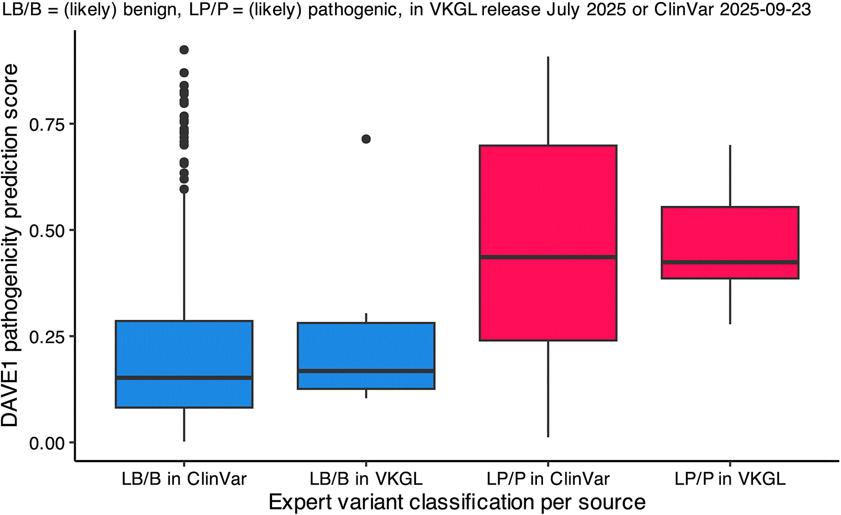
These variants were VUS in VKGL release April 2024 and have since been reclassified, categorized by classification label (LB/LP) and classification source.
To demonstrate how DAVE can guide interpretation of functional consequences, we performed a more detailed analysis of three VUS that were reclassified in the July 2025 VKGL data-sharing release compared to the April 2024 release or had a different classification in ClinVar. These analyses are enabled by DAVE’s ability to break down the final pathogenicity probability into separate feature contributions as SHAP values (Lundberg and Lee, 2017), capturing interactions unique to each prediction. SHAP values of contributing features, including their delta and unit, are shown in a cumulative plot per variant. ChimeraX (Meng et al., 2023) was used to visualize the predicted effects on protein structures.
2.3.1 WNT7B T111P
The variant in WNT7B NM_058238.3:c.331A>C causes the amino acid substitution Thr111Pro in the Wnt-7b protein. It was selected for having the highest absolute folding energy change of the variants with an updated classification in the VKGL dataset in Table 2. It is predicted to have a substantial increase in ∆G folding energy, see Figure 2A. A higher ∆G causes protein folding to occur less spontaneous, affecting correct folding and stability. Molecular visualization shows a change in the hydrogen bond integral in the secondary structures and their connecting elements (Figure 2B). The impact on protein structural integrity can be a good mechanistic explanation of the effect of this missense variant.
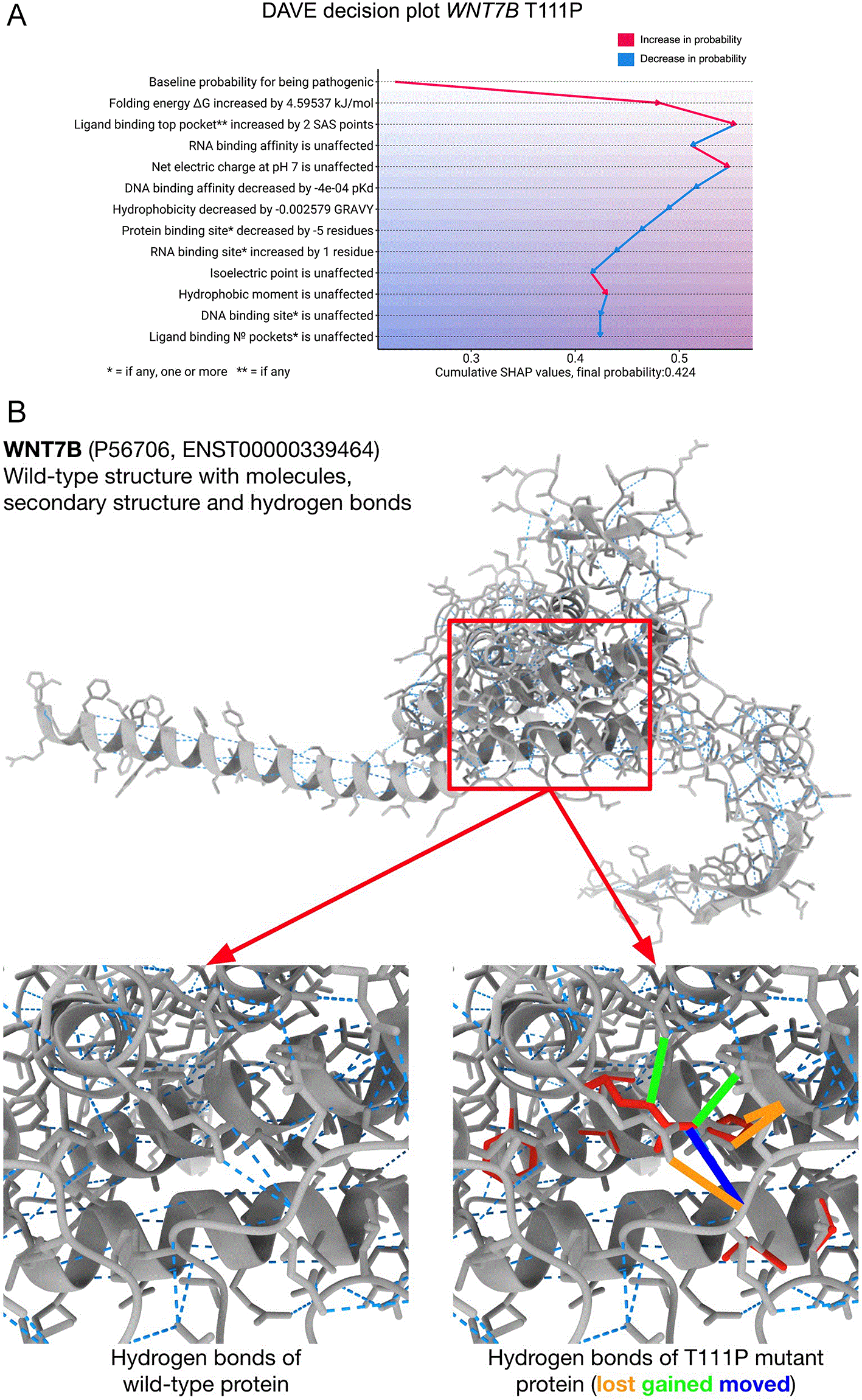
Features are on the y-axis, starting with a baseline pathogenicity probability, followed by contributing features ranked by their impact. Cumulative SHAP probability contributions are shown with arrows on the x-axis, ending on the final pathogenicity probability at the bottom. (B) Structural comparison of wild-type protein encoded by WNT7B to the variant protein with a T111P substitution. The complete protein wild-type structure is shown on top with, indicated with the red box are the affected region of the variant. Cropped images of the indicated region are shown at the bottom left and at the bottom right. The bottom left showing the wild-type structure. At the bottom right a cropped image of wild-type is shown with the T111P structure overlay in red. Hydrogen bonds are indicated with blue dotted lines with changes highlighted with solid lines.
2.3.2 SLCO2A1 G554R
The SLCO2A1 variant NM_005630.2:c.1660G>A, causes the amino acid substitution Gly554Arg in the solute carrier organic anion transporter family member 1A2 protein. It was selected for having the largest absolute change in solvent-accessible surface (SAS) points within its top-ranking ligand-binding pocket. Although currently still classified as a VUS in the VKGL dataset, it is reported as pathogenic in ClinVar (Xu et al., 2020). DAVE predicted the variant to be pathogenic, see Figure 3A. The predicted differences in the electrostatic surface potential of the ligand-binding pocket in the simulated structure of the G554R versus wild-type protein, are shown in Figure 3B. Moreover, recent functional characterization of variants in the transmembrane domain of SLCO2A1 replacing glycine with residues with larger side chains underscored this, as it revealed a loss of helical dynamics, accompanied by potential disruption of the helical packing (Xia et al., 2025).
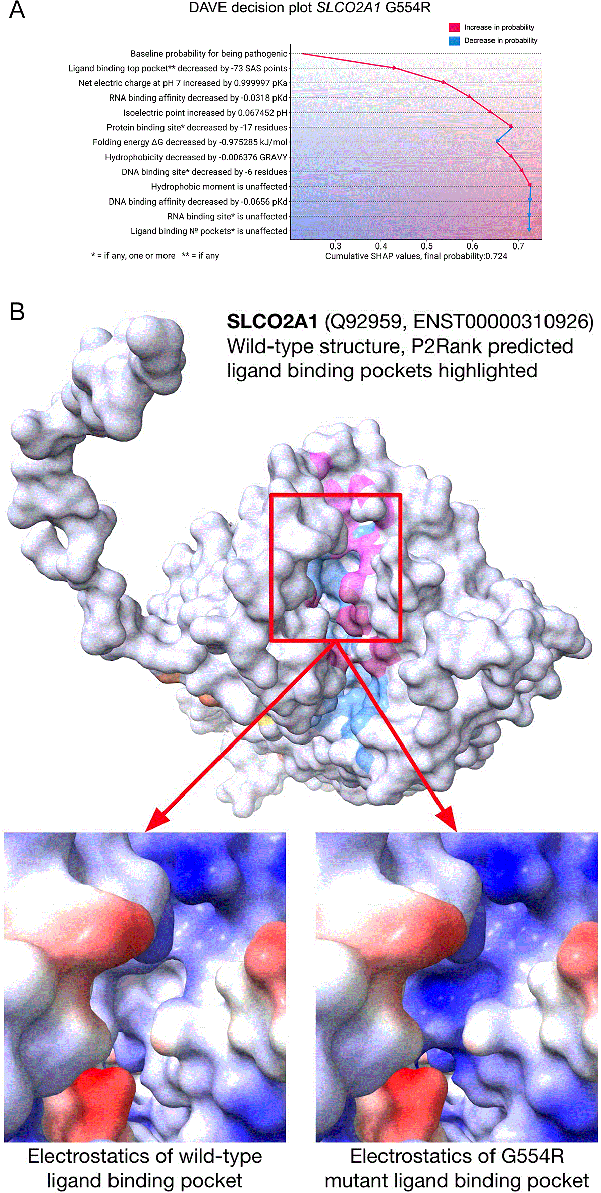
Features are on the y-axis, starting with a baseline pathogenicity probability, followed by contributing features ranked by their impact. Cumulative SHAP probability contributions are shown with arrows on the x-axis, ending on the final pathogenicity probability at the bottom. (B) Structural comparison of wild-type protein encoded by SLCO2A1 to the protein with the G554R substitution. Top: wild-type protein is displayed in white, with pink, light-blue, orange and yellow accents given to the predicted ligand binding pockets. The region of interest is indicated with the red box. Bottom: the Coulombic electrostatic surface potential is shown for region of interest, where red indicates negative potential and blue is positive potential.
2.3.3 NKX2–5 L153P
The variant NKX2–5 NM_004387.3: c.458T>C, causes the amino acid change, Leu153Pro in the homeobox Nkx-2.5 protein. It was selected for having largest absolute change in protein binding site residues. This variant is currently classified as VUS in the VKGL dataset while reported as pathogenic in ClinVar. DAVE predicts that this variant is pathogenic, shown in Figure 4A. The protein encoded by NKX2–5 is a transcription factor, and the substitution L153P, lies in an important homeodomain (AA 138–197) responsible for the binding of DNA as well as other transcription factors (Pradhan et al., 2012). Interestingly, despite the location of the variant, the most contributing feature to the predicted pathogenicity of this variant is not the change in binding site residues, but the change in folding energy ∆G. The predicted structural change of the L153P variant protein compared to the wild-type is visualized in Figure 4B.
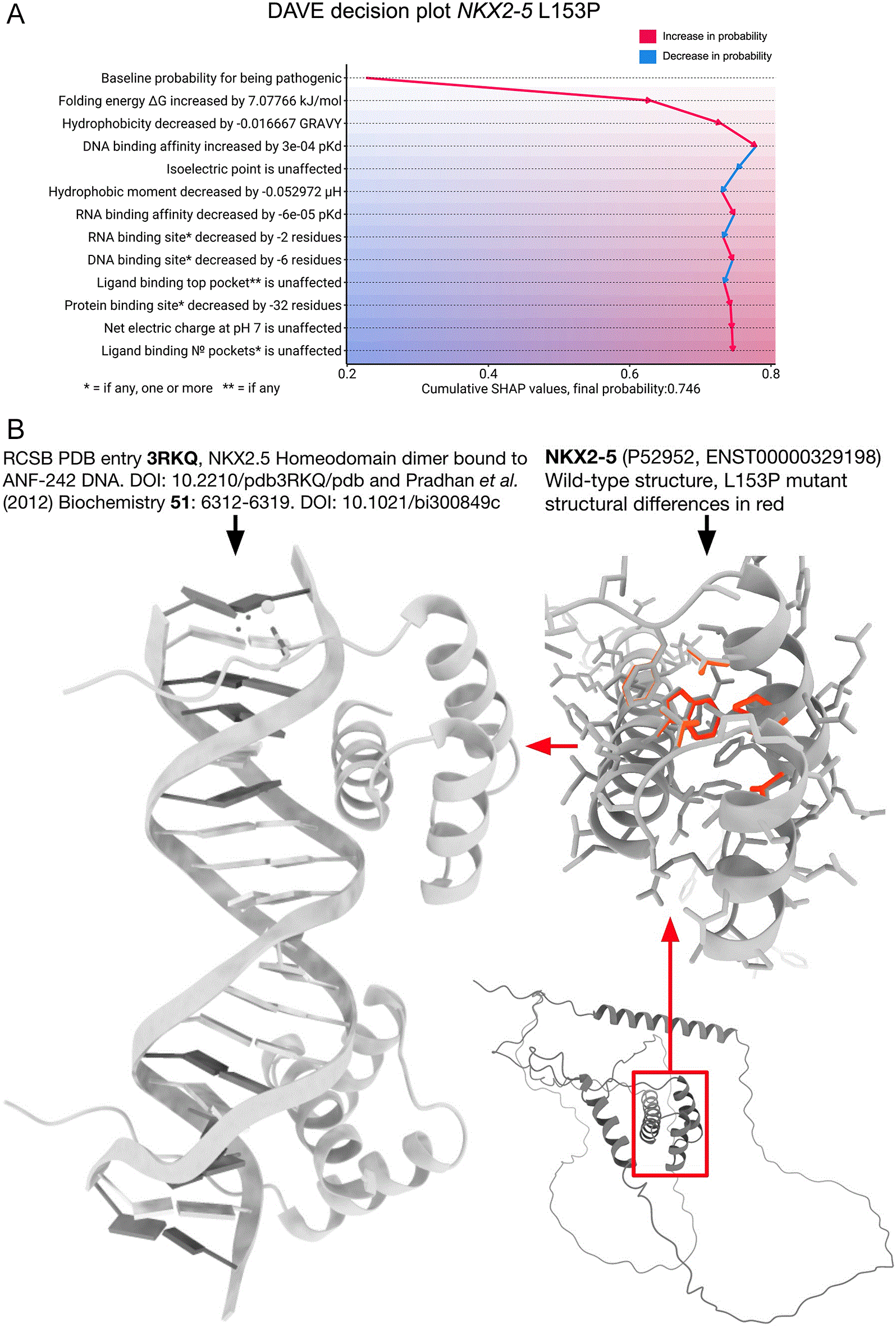
Features are on the y-axis, starting with a baseline pathogenicity probability, followed by contributing features ranked by their impact. Cumulative SHAP probability contributions are shown with arrows on the x-axis, ending on the final pathogenicity probability at the bottom. (B) Structural comparison of the protein with the L153P substitution to the wild-type protein encoded by the NKX2–5 gene. Left: Homeodomain (HD) interacting with ANF-242 DNA sequence. Right bottom: Highlighted in the red box, the location of the HD in the protein encoded by NKX2–5. Right top: HD where L153P structure changes are shown in red.
We developed MOLGENIS DAVE, a supervised learning model that predicts pathogenicity of missense variants and provides interpretable insights via contributions of functionally relevant features based on protein modeling. Unlike methods such as AlphaMissense (Chen et al., 2023) and REVEL (Ioannidis et al., 2016), DAVE prioritizes physical explainability over predictive power, providing complementary evidence for variant classification and guidance for targeted experimental validation of VUS either in dry or wet laboratory.
The selected features of DAVE represent multiple disease-causing mechanisms (Sen et al., 2022) and thus provide a starting point for interpretation. However, no single model can fully capture the complexity and unique biology of all proteins. Manual follow-up will remain essential to accurately determine the molecular consequences of prioritized variants. By integrating more diverse and context-dependent features, future versions of DAVE could better capture the nuanced mechanisms underlying variant effects, ultimately improving both accuracy and interpretability across a broader range of proteins and variant types. For instance, variants that have a gain-of-function or dominant-negative consequence have different, often milder, molecular mechanisms compared to loss-of-function variants (Gerasimavicius et al., 2022; Backwell and Marsh, 2022). Other examples of such effects include allostery, post-translational modifications (PTMs), toxicity, structural flexibility, folding rate, kinetic effects, aggregation propensity, membrane association, immunogenicity, and cooperativity.
Some AlphaFold structures are predicted with low confidence, particularly for disordered regions or poorly conserved domains (Luppino et al., 2025; Brotzakis et al., 2025). In these regions, predictive accuracy can be significantly lower, especially if models rely heavily on structural data (Kong et al., 2025). Another limitation of current models is that they represent the protein without their biological context, and that models larger than 2700 residues (<2% of all models) are chunked into smaller fragments due to computational limitations, making them unsuitable for meaningful predictions. To increase applicability across diverse protein types and missense effect types, structure confidence metrics and more biological context should be integrated to refine functional predictions. We expect that future versions of AlphaFold and similar efforts will produce unfragmented models for all proteins.
While DAVE could certainly be configured to run as part of DNA interpretation pipelines, currently it is not offered an easy to install standalone product. The reasons are the integration with external software, one of which used under academic personal license, as well as a prohibitive computational burden, averaging around 20 minutes per variant on commodity hardware. This hinders its applicability in clinical genetics workflows where rapid turnaround time is essential. For future versions, we will prioritize improving computational efficiency and ensuring permissive licensing of underlying methods.
In summary, DAVE combines predictive accuracy with meaningful information on how its predictions are made. By complementing numerical predictions with mechanistic insights, we demonstrate the potential to transform in silico variant effect predictions into interpretable molecularly grounded explanations. The insights provide a practical route to generate testable hypotheses for the resolution of VUS in functional follow-up studies, thereby bridging the gap between prediction, explanation, and clinical utility. Although DAVE captures multiple protein-related features, it does not currently account for variant effects through other mechanisms, such as altered gene expression or splicing. Consequently, pathogenicity arising from these processes is not assessed by the model. In such cases, the use of an ensemble of specialized tools covering complementary variant effects is recommended. This limitation is exemplified by our false negative prediction for CACNA1A Leu622Gln, see Table 2. This variant was classified in diagnostics as likely pathogenic due to a predicted impact on splicing, which explains why DAVE did not predict a significant pathogenic effect at the protein level. Looking ahead, we envision a framework that integrates DAVE with complementary evidence such as population frequency, conservation, inheritance, and phenotypes as well as specialized tools that cover effects on transcription and translation to systematically classify missense variants and reduce VUS. Integrating DAVE into pipelines such as MOLGENIS VIP (Maassen et al., 2025) would enable the inclusion of these complementary tools within a scalable and modular infrastructure for variant interpretation. Such an integrative approach enables large scale reanalysis efforts, to prioritize variants for experimental validation, and ultimately accelerate the route from variant discovery to informed clinical decision making.
The main resource is the fully de-identified and publicly accessible DNA variant classifications dataset collected in April 2024 by the Datashare working group of the nine genome diagnostic labs in the Netherlands, organized in the VKGL (Vereniging Klinisch Genetische Laboratoriumdiagnostiek) (Fokkema et al., 2019). We downloaded the GRCh38 liftover version from the MOLGENIS VIP (Maassen et al., 2025) resources. If variants within the same gene with the same protein change had differences in classification, they were marked as conflicting. The July 2025 version of the VKGL release, was collected similarly to the April 2024 release. For protein structures, we used pdb’s from the AlphaFold v4 release. AlphaFold structures are represented by UniProt and mappings to HGNC gene symbols are provided. The GRCh38 VCF file for ClinVar (Landrum et al., 2013) release 2025-09-23 was used as-is by matching on chromosome, position, reference and alternative allele.
The VKGL variant dataset was annotated with Ensembl VEP (McLaren et al., 2016) version 112. Based on protein localization data from the Protein Atlas (Uhlén et al., 2015) we randomly selected 1007 intracellular, 1028 membrane, and 692 secreted proteins for which we also had genomic variants in the dataset. This selection of proteins was used to subset the VKGL variants to balance the data on localization. Additionally, we annotated whether chaperones were involved in the folding of these proteins, for this we used a dataset of 194 manually curated chaperones (Shemesh et al., 2021) and expanded these genes with UniProt/SwissProt IDs using Ensembl BioMart. These known chaperones were connected to their interaction protein partners using BioGRID (Oughtred et al., 2020).
We applied the following five protein analysis tools on the selected variants, P2Rank (Krivák and Hoksza, 2018), FoldX 5 (Delgado et al., 2019), GLM-Score (Dias and Kolazckowski, 2015), Peptides R package (Osorio et al., 2014), and GeoNet (Han et al., 2024). Complete data processing was successful for 23,417 variants, of which the classifications, localizations, and chaperoned folding characteristics are shown in Table 3.
Each number represents a set of DNA variants randomly sampled from the VKGL release of April 2024, though equalized across protein localization and folding pathway. Benign includes ‘likely benign’ and ‘benign’, Pathogenic includes ’likely pathogenic’ and ’pathogenic’. VUS are variants of uncertain significance.
To obtain the most informative and independently contributing set of features, the set was minimized by removing correlations and selecting the most informative and explainable features. Among the remaining features, pairwise correlations were minimal, with the exception of correlation between‘Net electric charge at pH 7’ and ‘Isoelectric point’. Despite this, both features were deemed valuable to retain due to their individual relevance. The R function ‘cor’ from the stats (r-core) package was used to calculate the correlation of features as the Pearson correlation coefficient (PCC), shown in Figure 5.
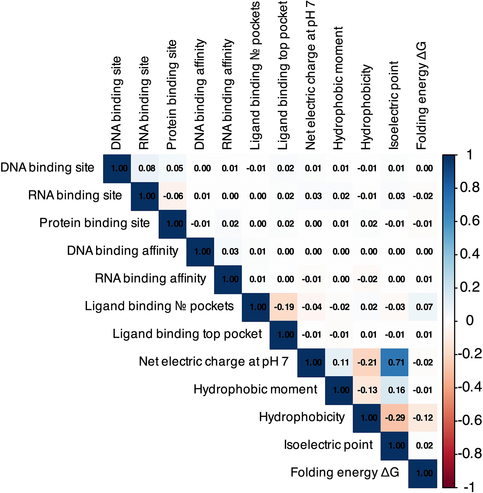
Correlation was calculated for every feature as the Pearson correlation coefficient.
A subset of the full annotated dataset, consisting of 12,048 variants classified as likely pathogenic (LP) or likely benign (LB), was split into training and testing sets using an 80/20 ratio. The DAVE model was implemented using the randomForest package in R (version 4.7–1.1) using the standard Random Forest algorithm with default parameters. To determine the contribution of each selected feature to individual predictions, SHAP values were calculated using the R package fastshap (version 0.1.1) with 10-fold Monte Carlo sampling. It is important to note that SHAP values do not correlate with feature values but instead capture feature contributions based on the interactions among all features uniquely for this prediction.
For binary classification, the VKGL April 2024 ‘test’ set was used to determine the optimal threshold with the R package cutpointr (version 1.1.2). From this, a threshold of 0.286 was established as the cutoff point using Youden’s J statistic expressed as:
J=TPTP+FN+TNTN+FP−1
TP = True Positive
TN = True Negative
FP = False Positive
FN = False Negative
To evaluate the utility of DAVE in the reclassification of the 11,221 variants of uncertain significance (VUS), we compared variant classifications between the April 2024 and July 2025 VKGL datasets. Additionally, we compared variants classified as VUS in the VKGL that had a B/LB or LP/P classification in ClinVar (September 23, 2025, with at least a one-star review status and no conflicting classifications) with the predictions of DAVE. For variant specific structural comparison, we substituted wild-type amino acid with the variant in the AlphaFold (Jumper et al., 2021) structure, PDBs using FoldX5 (Delgado et al., 2019) with the BuildModel and RepairPDB command. These structures were then visualized with ChimeraX (Meng et al., 2023) and compared with wild-type structures.
All code used in the training and testing of the model, as well as the code to generate the figures is available as free and open-source software:
Project name: MOLGENIS DAVE.
Project homepage: https://github.com/molgenis/dave
Availability: Archived on Zenodo ‘DAVE codebase and resources’ (DOI: 10.5281/zenodo.20505823) and available on https://github.com/molgenis/dave (van der Velde, 2026).
Operating system(s): Ubuntu Linux or Windows using Windows Subsystem for Linux (WSL).
Programming language(s): R (version 4.3.3), Java (version 11).
License: code is licensed under LGPL-v3/all non-code content under CC-BY-4.0.
Other requirements:
Not applicable.
Not applicable.
We would like to thank our colleagues at Genomics Coordination Center for engineering support and access to the UMCG high-performance compute cluster. We are grateful to our colleagues from all Dutch genome diagnostic laboratories for their continuing efforts to share variant classifications through the VKGL Datashare working group. We would also like to thank our previous and current heads of department Prof. Nine Knoers and Prof. Anke-Hilse Maitland-van der Zee as well as the UMCG Board of Directors, in particular Prof. Stephanie Klein Nagelvoort Schuit and Prof. Wiro Niessen for their encouragement and support to develop and use new AI methods at the University Medical Center Groningen. Lastly, we would like to express our gratitude to the exquAIro AI bootcamp organization and board members Prof. Gerard Koppelman, Dr. Martin Smit, Ilya Petoukhov, Dr. Marnix Bügel, as well as Geerte Koster of REWIRE and Dr. Kai Yu Ma of UMCG for their training, coaching and valuable input.
This research was supported by the ERDERA project (grant agreement No. 101156595), which has received funding from the European Union’s Horizon Europe research and innovation programme, and The Netherlands Organisation for Scientific Research NWO under VIDI grant number 917.164.455.
The funders had no role in study design, data collection and analysis, decision to publish, or preparation of the manuscript.
© 2026 Niemeijer T et al. This is an open access article distributed under the terms of the Creative Commons Attribution License, which permits unrestricted use, distribution, and reproduction in any medium, provided the original work is properly cited.
Current Reviewer Status:
AWAITING PEER REVIEW
AWAITING PEER REVIEW
?
Key to Reviewer Statuses VIEW HIDE
ApprovedThe paper is scientifically sound in its current form and only minor, if any, improvements are suggested
Approved with reservations A number of small changes, sometimes more significant revisions are required to address specific details and improve the papers academic merit.
Not approvedFundamental flaws in the paper seriously undermine the findings and conclusions
Open Peer Review
AWAITING PEER REVIEW
Sign up for content alerts