Просмотр новости
Найдите то, что Вас интересует
 Спутники
 РН
РН SpaceX
 Роскосмос
 NASA
NASA
СпутникиРНSpaceXРоскосмосNASAPosterior reversible encephalopathy syndrome (PRES) is an acute neurotoxic condition characterized by headache, seizures, altered mental status, and visual disturbance in association with vasogenic edema on neuroimaging.1,2 It is most frequently reported in the context of severe hypertension, renal disease, cytotoxic or immunosuppressive therapy, and (pre)eclampsia.2,5 We report a 31-year-old primigravida at 26 weeks’ gestation following in vitro fertilization (IVF) twin pregnancy, who presented with abrupt, painless, bilateral loss of vision as the only neurological manifestation of preeclampsia-associated PRES. On admission, blood pressure was mildly elevated at 143/94 mmHg, and systemic and neurological examinations were otherwise unremarkable apart from brisk reflexes. Laboratory investigations were consistent with preeclampsia, and brain MRI demonstrated right-predominant parieto-occipital cortical signal changes in keeping with PRES.8,11 Stroke, cerebral venous sinus thrombosis, and primary ophthalmic causes were excluded by clinical assessment and imaging. The patient was treated with antihypertensive therapy, magnesium sulfate, and expedited delivery by caesarean section, resulting in complete visual recovery within 48 hours and favorable early maternal-neonatal outcomes. This case highlights an uncommon PRES phenotype: isolated reversible bilateral blindness in the setting of only mild hypertension, IVF twin pregnancy, and asymmetric imaging, underscoring the importance of maintaining a high index of suspicion for PRES in preeclamptic patients presenting with acute visual loss.
Posterior reversible encephalopathy syndrome (PRES), first described by Hinchey et al. in 1996, is a clinico-radiological entity characterized by acute neurological symptoms and reversible subcortical vasogenic edema predominantly affecting the parieto-occipital regions.1 Previously termed reversible posterior leukoencephalopathy syndrome, PRES lies at the intersection of neurology and systemic disease, most often arising in the context of acute blood pressure fluctuations, endothelial dysfunction, or exposure to cytotoxic and immunosuppressive agents.2,5
Clinically, PRES typically presents with a constellation of headache, seizures, confusion or decreased level of consciousness, visual disturbances, and focal neurological deficits.2,6,8 Although visual impairment is common, complete, abrupt, bilateral visual loss (cortical blindness) without accompanying seizures or encephalopathy is distinctly uncommon.3,4 Preeclampsia and eclampsia are well-recognized precipitants of PRES, generally in association with severe or fluctuating hypertension.2,5,9
The pathophysiology of PRES is incompletely understood. A widely accepted hypothesis proposes that severe hypertension overwhelms cerebral autoregulation, resulting in hyperperfusion, endothelial injury, and vasogenic edema.5 An alternative model suggests that hypertension induces cerebral vasoconstriction, hypoperfusion, and subsequent ischemia, reflecting a more ischemic mechanism.5 The coexistence of cases with normal or only mildly elevated blood pressure suggests that endothelial dysfunction and breakdown of the blood-brain barrier may be central to the syndrome, particularly in conditions such as preeclampsia.5
Common etiologies associated with PRES include renal disease, autoimmune disorders, chemotherapy and immunosuppressive therapy, preeclampsia/eclampsia, and sepsis or septic shock.2,8 Proposed diagnostic criteria include acute onset of neurological symptoms, compatible neuroimaging demonstrating vasogenic edema, and reversibility of clinical and/or radiological findings.2,6
We describe an unusual case of preeclampsia-associated PRES in an IVF twin pregnancy, presenting solely with acute bilateral vision loss in the setting of mild hypertension and right-predominant parieto-occipital involvement on MRI. This case adds to the limited literature on atypical PRES presentations in pregnancy and emphasizes the need for early MRI and consideration of PRES even when blood pressure is not severely elevated.
A previously healthy 31-year-old woman, primigravida at 26 weeks’ gestation, presented to the emergency department with sudden, painless, bilateral loss of vision. The pregnancy resulted from in vitro fertilization and was a dichorionic twin gestation that had been uncomplicated until presentation. Her antenatal care had been regular, and she had no known history of chronic hypertension, diabetes, renal disease, or autoimmune disorders. Current medications included oral and injectable progesterone, low-dose aspirin, and routine antenatal supplements. Family history was notable for maternal hypertension and diabetes.
On the morning of presentation, the patient awoke with blurry vision that progressed over a short period to complete loss of vision following a brief period of rest. She denied eye pain, diplopia, focal weakness, speech disturbance, seizures, or altered consciousness. She reported a mild headache and nasal congestion during the preceding four days but no fever or systemic symptoms.
On arrival, she was alert, fully oriented, and speaking calmly. Vital signs showed blood pressure of 143/94 mmHg, heart rate 94 beats per minute, respiratory rate 20 breaths per minute, and oxygen saturation 98% on room air. Cardiovascular, respiratory, and abdominal examinations were unremarkable. Neurological examination demonstrated intact cranial nerves, normal motor strength, and preserved sensation. Deep tendon reflexes were brisk throughout, with no clonus, and plantar responses were flexor. Visual acuity could not be measured due to complete functional blindness, but pupils were equal and reactive, and there was no relative afferent pupillary defect.
Initial laboratory investigations revealed 3+ proteinuria, elevated lactate dehydrogenase and uric acid, and reduced total protein and albumin, consistent with preeclampsia. Renal and liver function tests, full blood count, and coagulation profile were otherwise within normal limits. Table 1 summarizes the laboratory values at admission.
Given the acute onset of bilateral visual loss in a pregnant patient, a differential diagnosis included; Ischemic or hemorrhagic stroke involving the posterior circulation, Cerebral venous sinus thrombosis (CVST), Optic neuritis or ischemic optic neuropathy, Retinal pathology (e.g., central retinal artery or vein occlusion, serous retinal detachment) and PRES associated with preeclampsia.
An urgent ophthalmology assessment found normal anterior segment and fundus examination, with no evidence of optic disc swelling, retinal hemorrhages, vascular occlusion, or retinal detachment, arguing against primary ocular or optic nerve pathology. Neurological evaluation yielded a National Institutes of Health Stroke Scale (NIHSS) score of 3, attributed solely to her visual deficit, with no motor, sensory, or speech impairment. The absence of focal neurological signs, normal coagulation studies, and lack of risk factors for thrombophilia made acute ischemic stroke and CVST less likely, although imaging was required to exclude these entities definitively.
Brain Magnetic Resolution Imaging (MRI) with diffusion-weighted imaging (DWI) showed right parieto-occipital cortical areas of abnormal signal, with findings consistent with vasogenic edema in a pattern typical for PRES. The basal ganglia, lateral thalamus, and selected cortical gyri showed increased signal intensity, without evidence of restricted diffusion suggestive of acute infarction. There was no imaging evidence of cerebral venous sinus thrombosis or intracranial hemorrhage. Figure 1 illustrates the axial images of the brain, demonstrating hyperintense areas in the right parieto-occipital region.
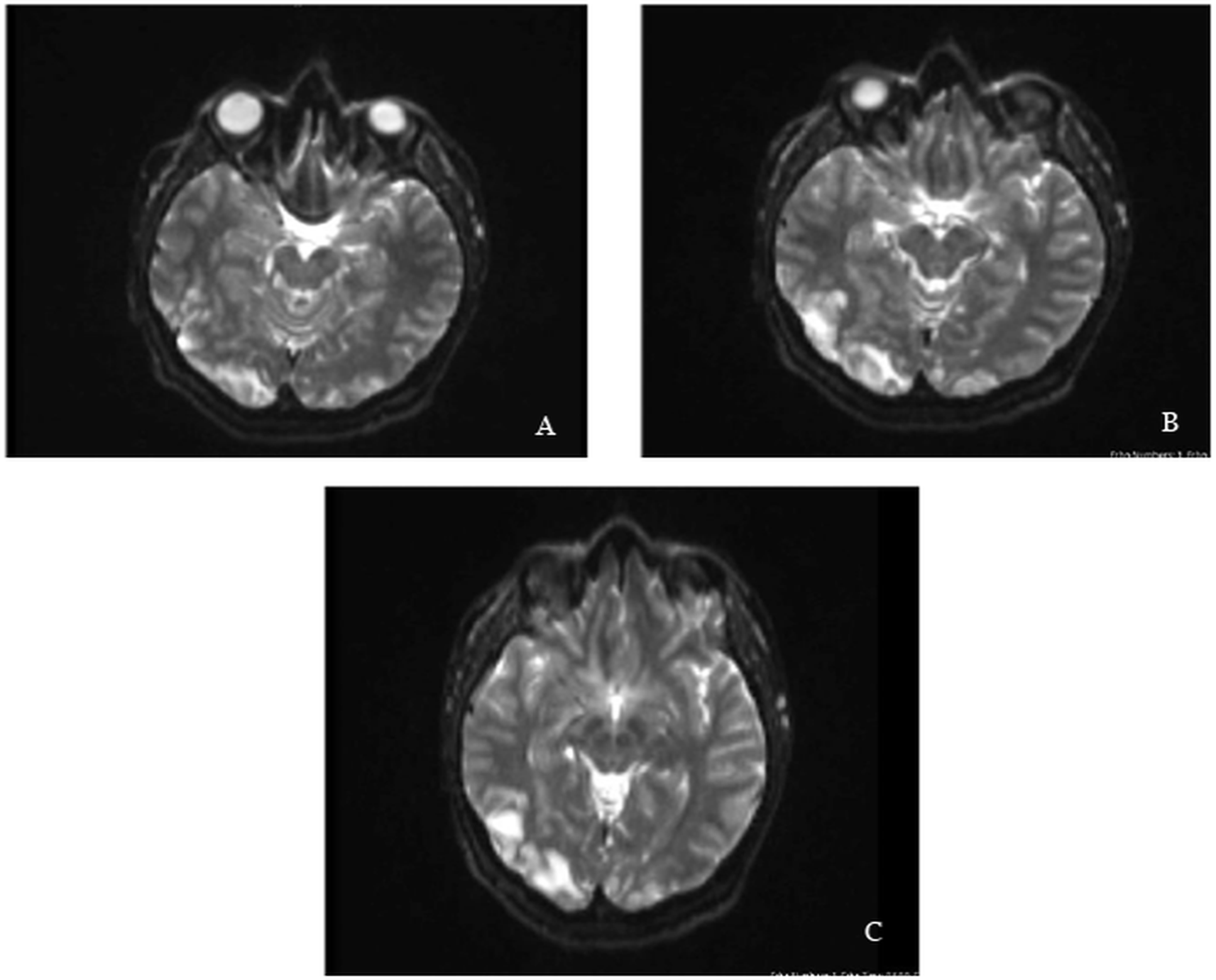
Axial images of the brain demonstrate hyperintense areas on the right parieto-occipital regions. The basal ganglia, lateral thalamus, and some cortical gyri show increased signal intensity on DWI.
Based on the clinical context of preeclampsia, acute bilateral cortical visual loss, and MRI findings of predominantly right parieto-occipital vasogenic edema, a diagnosis of PRES secondary to preeclampsia was established.
Management focused on controlling blood pressure, preventing seizures, and treating the underlying preeclampsia. The patient was initiated on intravenous labetalol and magnesium sulfate according to institutional preeclampsia protocols. The obstetrics team confirmed fetal viability for both twins and, after multidisciplinary discussion with neurology and anesthesiology, recommended expedited delivery as definitive treatment for preeclampsia and PRES. She underwent an uneventful emergency caesarean section. Twin A and Twin B were delivered with birth weights of 0.95 kg and 0.83 kg, respectively, and Apgar scores of 4 and 8 for Twin A and 5 and 9 for Twin B at 1 and 5 minutes. Both neonates developed respiratory distress syndrome and required admission to the neonatal intensive care unit (NICU), where they received oxygen therapy, surfactant, and antibiotics.
Within 48 hours of delivery, the patient experienced complete recovery of visual function, with restoration of normal visual acuity and visual fields on clinical examination. Headache resolved, and she reported no further neurological symptoms. By day 3, her blood pressure had improved and was controlled on oral antihypertensive therapy. She was transferred from the high dependency unit to a general ward and subsequently discharged home in stable condition with instructions for regular blood pressure monitoring and follow-up in the obstetric and neurology clinics.
On early outpatient follow-up at 2 weeks post-discharge, she remained normotensive on oral antihypertensive medication with no recurrent visual or neurological complaints. Ophthalmology review confirmed normal ocular examination without residual deficits, with full restoration of visual fields and acuity. Blood pressure readings at home were within normal limits, and laboratory investigations showed resolution of proteinuria and normalization of serum albumin and uric acid levels.
The twins remained in the NICU for 6 weeks for ongoing management of prematurity-related complications including respiratory distress syndrome and feeding difficulties. Both neonates showed gradual respiratory improvement with weaning from oxygen support by 4 weeks of age. They were discharged home in stable condition at corrected gestational age of 32 weeks, with appropriate weight gain and feeding established. Early developmental assessments at three months corrected age were within normal limits for both infants.
Posterior reversible encephalopathy syndrome is a rare but potentially reversible neurotoxic condition characterized by acute neurological symptoms and distinctive radiological findings. It was initially described in 1996 by Hinchey et al. in a seminal case series of 15 patients who presented with headache, seizures, altered mental status, and visual disturbances in the setting of various acute illnesses.1 In that cohort, seven patients were receiving immunosuppressive therapy, four had hypertensive encephalopathy associated with renal disease, three had eclampsia, and one was on interferon, illustrating the broad range of systemic conditions that can precipitate PRES.1 Their work established the concept of a reversible posterior leukoencephalopathy with characteristic imaging features, which has since been refined into the entity now known as PRES.1
Conditions associated with PRES broadly fall into two overlapping categories: states of severe or fluctuating hypertension with cerebral hyperperfusion, and states characterized by endothelial dysfunction that impair cerebral autoregulation.2 The former includes hypertensive emergencies and acute renal failure, whereas the latter encompasses preeclampsia and eclampsia, solid organ transplantation, sepsis and septic shock, cytotoxic chemotherapy, and a range of autoimmune diseases such as systemic lupus erythematosus, systemic sclerosis, granulomatosis with polyangiitis, and polyarteritis nodosa.2,8 Miscellaneous associations such as electrolyte disturbances, Guillain–Barré syndrome, tumour lysis syndrome, and certain antibiotics have also been reported, highlighting that PRES is a final common pathway of diverse vascular and endothelial insults.7,8
Published case reports reinforce this heterogeneity. One report described development of PRES after only one day of metronidazole therapy for Clostridioides difficile infection, where the patient progressed from mild headache and dizziness to altered mental status and a left-sided homonymous hemianopia.7 Like our patient, that case did not exhibit markedly elevated blood pressure, emphasizing that PRES can occur outside the classic severe hypertensive context.7 However, our case differs in two important respects: there were no changes in mental status at any point, and the visual impairment manifested as complete bilateral cortical blindness rather than a unilateral visual field deficit.7 This contrast underscores the wide clinical spectrum of PRES and illustrates how isolated visual loss can be the dominant or only neurological sign.
The clinical presentation of PRES is highly variable, ranging from headache, nausea and vomiting to seizures, altered level of consciousness, visual disturbances, and focal deficits including hemiparesis.6,8 Seizures are often generalized tonic–clonic and may be preceded by visual auras or other focal symptoms.6 Post-ictal confusion of varying duration is common, and deep tendon reflexes are frequently exaggerated, with extensor plantar responses in some patients.6 In our case, the absence of seizures, preserved consciousness, and normal motor and sensory examination made the presentation atypically “pure visual”, which risks misclassification as an isolated ophthalmic or functional visual disorder if neuroimaging is delayed.
Cortical blindness is a recognized manifestation of preeclampsia-associated PRES, but most published cases describe severe hypertension, seizures, and encephalopathy in addition to visual loss.9 A smaller subset of reports document PRES in normotensive or mildly hypertensive patients, yet even in these, seizures or confusion are usually present.10 The combination of preeclampsia, only mildly elevated blood pressure, intact mental status, and isolated reversible cortical blindness therefore appears to be uncommon in the literature.3,4 Our case may be particularly informative for clinicians evaluating sudden visual loss in the antenatal or peripartum period, where attention often focuses on retinal or optic nerve pathology and cerebrovascular events.
The pathophysiology of PRES remains incompletely understood. The traditional hyperperfusion model proposes that abrupt, severe hypertension exceeds the limits of cerebral autoregulation, leading to arteriolar dilatation, hyperperfusion, capillary leakage, and vasogenic edema, most prominently in the posterior circulation where sympathetic innervation is relatively sparse.5 This model aligns well with cases of PRES occurring in hypertensive emergencies and renal crises.5 However, the observation that about one-third of patients with PRES present with normal or only mildly elevated blood pressure challenges a purely pressure-driven mechanism.5,10 These cases support an alternative paradigm centred on endothelial dysfunction and blood–brain barrier breakdown, driven by circulating toxins, cytokines, or immune complexes rather than absolute pressure levels.5
Preeclampsia is paradigmatic for this endothelial model. Although clinically defined by hypertension and proteinuria, it is fundamentally a systemic endothelial disorder characterized by abnormal placentation, anti-angiogenic factor release, and widespread microvascular injury.5 In our patient, the mean arterial pressure at presentation (approximately 110 mmHg) did not exceed the usual upper limits of autoregulation, yet she developed radiological features of PRES. This supports the hypothesis that, in preeclampsia-associated PRES, endothelial injury and impaired autoregulatory capacity may be sufficient to precipitate vasogenic edema even in the absence of severe hypertension.5 The additional presence of an IVF twin pregnancy may further amplify endothelial stress and hemodynamic load, although data in this specific subgroup remain limited.
Diagnosing PRES can be challenging because there are no universally accepted diagnostic criteria, and its features overlap with other acute neurovascular disorders.2,12,13 Fugate and colleagues proposed pragmatic criteria that are widely used in practice: acute onset neurological symptoms, neuroimaging abnormalities consistent with vasogenic edema, and reversibility of clinical and/or radiological findings over time.13 Importantly, PRES remains a diagnosis of exclusion; alternative causes such as ischemic or hemorrhagic stroke, cerebral venous sinus thrombosis, encephalitis, and neoplastic or metastatic lesions must be considered and ruled out based on clinical, laboratory, and imaging data.12 In our case, the normal coagulation profile, lack of focal neurological deficits, and MRI pattern of vasogenic rather than cytotoxic edema argued strongly against stroke or venous thrombosis.
Imaging plays a central role in confirming PRES. In the acute setting, CT is often the first modality obtained and may show areas of white-matter hypoattenuation in regions typically affected by PRES, such as the parietal and occipital lobes.8 However, CT is relatively insensitive and may be normal, especially early in the course.8 MRI offers superior sensitivity and anatomical detail, usually demonstrating bilateral, symmetric hyperintensities on T2-weighted and FLAIR sequences in the parieto-occipital regions, sometimes extending to the frontal lobes, temporal–occipital junction, or cerebellum.8,11 As edema progresses, lesions can become confluent.8 Diffusion-weighted imaging typically shows vasogenic rather than cytotoxic edema, helping to distinguish PRES from acute infarction.8,11 In our patient, MRI revealed predominantly right-sided parieto-occipital involvement with associated signal changes in the basal ganglia and thalamus, representing an asymmetric but recognized pattern within the PRES spectrum.8,11
Additional investigations can support the diagnosis or exclude mimics. Electroencephalography is useful for detecting subclinical seizures or non-convulsive status epilepticus, which may present with reduced responsiveness or fluctuating mental status.2 Lumbar puncture and cerebrospinal fluid analysis can help rule out infectious or inflammatory encephalitis and leptomeningeal metastasis in appropriate clinical contexts.2 Serum studies, including autoimmune panels, metabolic screens, and drug levels, may identify underlying triggers or co-morbidities.2 In our patient, the absence of encephalopathy, focal deficits, or systemic signs of infection reduced the yield of EEG and CSF examination, so management centred on neuroimaging findings and the obstetric context.
Management of PRES focuses on rapid recognition, treatment of the underlying cause, and prevention of complications such as seizures and intracranial hemorrhage.2,14 Blood pressure control with carefully titrated antihypertensives is essential, avoiding both severe hypertension and rapid over-correction that could compromise cerebral perfusion.14 In pregnancy-related cases, magnesium sulfate serves both as seizure prophylaxis and as standard therapy for preeclampsia.9 When PRES is linked to immunosuppressive or cytotoxic therapy, dose reduction or discontinuation of the offending agent is often required.14 In our case, definitive treatment of the precipitating condition was achieved through expedited caesarean delivery, consistent with established principles that delivery is the only curative therapy for preeclampsia.9
The prognosis of PRES is generally favorable when recognized early and managed appropriately, with most patients experiencing substantial or complete recovery of neurological function.2,14,15 Nevertheless, delayed diagnosis, persistent uncontrolled hypertension, or extensive cytotoxic edema can lead to permanent deficits or even death.15 Our patient’s rapid visual recovery within 48 hours and sustained normal neurological and ophthalmological status on follow-up underscore the potential for full reversibility when multidisciplinary care is promptly instituted. For the neonates, the course was dominated by prematurity-related complications rather than direct effects of PRES, but early NICU involvement allowed a favorable short-term outcome.
This case highlights several key learning points. First, PRES should be considered in preeclamptic patients with acute visual symptoms, even in the absence of severe hypertension, seizures, or altered mental status. Second, MRI is essential for differentiating PRES from stroke, CVST, and other causes of cortical blindness, particularly when clinical findings are subtle. Third, IVF twin pregnancy and underlying endothelial vulnerability may predispose to atypical PRES phenotypes, including asymmetric lesions and isolated cortical blindness.
This case illustrates an uncommon presentation of preeclampsia-associated PRES manifesting as isolated, reversible bilateral cortical blindness in the setting of only mildly elevated blood pressure, IVF twin pregnancy, and predominantly right-sided parieto-occipital involvement on MRI. It emphasizes the need for a high index of suspicion for PRES in pregnant patients with acute visual loss, timely MRI to confirm the diagnosis, and prompt multidisciplinary management with blood pressure control, seizure prophylaxis, and expedited delivery when indicated. Early recognition and coordinated care can result in complete neurological recovery for the mother while optimizing outcomes for preterm neonates.
The Medical Research Center at Hamad Medical Corporation in Qatar granted approval for the publication of this case report.
Written informed consent was obtained from the patient for publication of details of their medical case and any accompanying images.