Просмотр новости
Найдите то, что Вас интересует
 Спутники
 РН
РН SpaceX
 Роскосмос
 NASA
NASA
СпутникиРНSpaceXРоскосмосNASABackground Calotropis gigantea latex is an uncommon cause of chemical ocular injury that can induce corneal epithelial damage, stromal edema, anterior uveitis, and endothelial dysfunction. Although most reported cases achieve favorable clinical and visual recovery with conservative treatment, objective evidence of persistent endothelial damage using multimodal ocular imaging remains limited. We report a case highlighting the value of anterior segment optical coherence tomography (AS-OCT) and specular microscopy in assessing acute corneal changes and long-term endothelial injury. Case Presentation A 72-year-old woman presented with a 4-day history of painful visual loss, redness, watering, photophobia, and inability to open the right eye after accidental exposure to Calotropis gigantea latex. Examination revealed hand movement vision, elevated intraocular pressure (25 mmHg), a large corneal epithelial defect (7 × 9 mm), diffuse stromal edema, Descemet membrane folds, and 4+ anterior chamber cells with a 0.5-mm hypopyon. Initial microbiological investigations were negative. AS-OCT demonstrated marked corneal thickening (approximately 940 μm) with diffuse stromal hyperreflectivity. Following medical treatment, the epithelial defect healed, inflammation resolved, and best-corrected visual acuity (BCVA) improved to 6/6 within four weeks. However, at one-year follow-up, specular microscopy demonstrated persistent severe endothelial cell loss (648 cells/mm2) with marked polymegathism and pleomorphism despite a clinically clear cornea. Conclusion Calotropis gigantea latex induced chemical ocular injury may result in irreversible endothelial damage despite complete clinical recovery. Multimodal ocular imaging, particularly AS-OCT and specular microscopy, is valuable for documenting acute corneal changes and detecting persistent subclinical endothelial compromise, supporting its role in long-term prognostic assessment.

Pandey S, Sitaula S, Shrestha S and Chaudhary M. Case Report: Multimodal ocular imaging of persistent corneal endothelial cell loss following Calotropis gigantea latex induced chemical ocular injury: a 1-year follow-up [version 1; peer review: awaiting peer review]. F1000Research 2026, 15:1357 (https://doi.org/10.12688/f1000research.188208.1)
Case Report
[version 1; peer review: awaiting peer review]
https://orcid.org/0009-0001-4856-4126
1, Sanjeeta Sitaula1, Sauravi Shresthahttps://orcid.org/0000-0002-5558-6358
1, Meenu Chaudhary1https://orcid.org/0009-0001-4856-4126
1, Sanjeeta Sitaula1, Sauravi Shresthahttps://orcid.org/0000-0002-5558-6358
1, Meenu Chaudhary11 Department of Ophthalmology, B.P. Koirala Lions Centre for Ophthalmic Studies, Maharajgunj Medical Campus, Institute of Medicine, Tribhuvan University, Kathmandu, Nepal
Sagar Pandey
Roles: Conceptualization, Data Curation, Investigation, Methodology, Writing – Original Draft Preparation
Sanjeeta Sitaula
Roles: Formal Analysis, Supervision, Validation, Writing – Original Draft Preparation, Writing – Review & Editing
Sauravi Shrestha
Roles: Data Curation, Investigation, Resources, Writing – Review & Editing
Meenu Chaudhary
Roles: Conceptualization, Project Administration, Supervision, Writing – Review & Editing
OPEN PEER REVIEW
REVIEWER STATUS AWAITING PEER REVIEW
Calotropis gigantea latex is an uncommon cause of chemical ocular injury that can induce corneal epithelial damage, stromal edema, anterior uveitis, and endothelial dysfunction. Although most reported cases achieve favorable clinical and visual recovery with conservative treatment, objective evidence of persistent endothelial damage using multimodal ocular imaging remains limited. We report a case highlighting the value of anterior segment optical coherence tomography (AS-OCT) and specular microscopy in assessing acute corneal changes and long-term endothelial injury.
Case PresentationA 72-year-old woman presented with a 4-day history of painful visual loss, redness, watering, photophobia, and inability to open the right eye after accidental exposure to Calotropis gigantea latex. Examination revealed hand movement vision, elevated intraocular pressure (25 mmHg), a large corneal epithelial defect (7 × 9 mm), diffuse stromal edema, Descemet membrane folds, and 4+ anterior chamber cells with a 0.5-mm hypopyon. Initial microbiological investigations were negative. AS-OCT demonstrated marked corneal thickening (approximately 940 μm) with diffuse stromal hyperreflectivity. Following medical treatment, the epithelial defect healed, inflammation resolved, and best-corrected visual acuity (BCVA) improved to 6/6 within four weeks. However, at one-year follow-up, specular microscopy demonstrated persistent severe endothelial cell loss (648 cells/mm2) with marked polymegathism and pleomorphism despite a clinically clear cornea.
ConclusionCalotropis gigantea latex induced chemical ocular injury may result in irreversible endothelial damage despite complete clinical recovery. Multimodal ocular imaging, particularly AS-OCT and specular microscopy, is valuable for documenting acute corneal changes and detecting persistent subclinical endothelial compromise, supporting its role in long-term prognostic assessment.
Calotropis gigantea, chemical ocular injury, corneal endothelial cell density, specular microscopy, anterior segment optical coherence tomography, Imaging, Infection, Ocular Surface, Anterior Uveitis, Keratitis
Corresponding author: Sagar Pandey Competing interests: No competing interests were disclosed.
Grant information: The author(s) declared that no grants were involved in supporting this work.
Copyright: © 2026 Pandey S et al. This is an open access article distributed under the terms of the Creative Commons Attribution License, which permits unrestricted use, distribution, and reproduction in any medium, provided the original work is properly cited. How to cite: Pandey S, Sitaula S, Shrestha S and Chaudhary M. Case Report: Multimodal ocular imaging of persistent corneal endothelial cell loss following Calotropis gigantea latex induced chemical ocular injury: a 1-year follow-up [version 1; peer review: awaiting peer review]. F1000Research 2026, 15:1357 (https://doi.org/10.12688/f1000research.188208.1) First published: 12 Aug 2026, 15:1357 (https://doi.org/10.12688/f1000research.188208.1) Latest published: 12 Aug 2026, 15:1357 (https://doi.org/10.12688/f1000research.188208.1)
Chemical ocular injuries are ophthalmic emergencies that can result in permanent visual impairment if not managed promptly.1,2 Although alkali and acid burns are the most common causes, plant-derived ocular toxicity remains an important yet underrecognized cause of corneal injury, particularly in regions where Calotropis species are widely used for traditional medicine and religious practices.3–11 The milky latex of Calotropis gigantea contains biologically active compounds capable of inducing epithelial toxicity, stromal edema, anterior uveitis, and endothelial dysfunction.3–7 Most reported cases demonstrate favorable clinical recovery following medical treatment; however, objective evidence of long-term endothelial damage is limited. Advances in multimodal ocular imaging, including anterior segment optical coherence tomography (AS-OCT) and specular microscopy, permit detailed assessment of corneal structural changes and endothelial integrity.12,13 We report a case of Calotropis gigantea latex induced chemical ocular injury in which multimodal imaging documented persistent severe endothelial cell loss despite complete anatomical and visual recovery.
A 72-year-old woman presented with a 4-day history of painful diminution of vision, redness, watering, photophobia, and inability to open her right eye following accidental exposure to the latex of Calotropis gigantea while applying the plant over sore areas of her body as a traditional remedy ( Figure 1A). She immediately irrigated the affected right eye with tap water; however, her symptoms progressively worsened. On examination, best-corrected visual acuity (BCVA) was hand movements in the affected right eye and 6/6 in the normal left eye. Intraocular pressure (IOP) measured 25 mmHg in the affected right eye and 17 mmHg in the left eye. Slit-lamp examination of the affected right eye demonstrated diffuse bulbar and circumciliary conjunctival congestion with a papillary conjunctival reaction. A large corneal epithelial defect measuring approximately 7 × 9 mm involved most of the cornea while sparing the limbus with no evidence of limbal ischemia. Multiple epithelial microbullae, diffuse stromal edema, and Descemet membrane folds were present. The anterior chamber was of normal depth and showed 4+ anterior chamber cells according to the Standardization of Uveitis Nomenclature (SUN) grading system14 with non-mobile inferior hypopyon measuring approximately 0.5 mm (Figure 1B). Fluorescein staining under cobalt blue illumination demonstrated intense uptake corresponding to the epithelial defect with well-defined margins ( Figure 1C) Due to dense corneal edema and anterior chamber reaction, the posterior segment could not be visualized. B-scan ultrasonography demonstrated a normal posterior segment. Anterior segment optical coherence tomography (AS-OCT) showed marked corneal thickening approximately of 940 μm with diffuse stromal hyperreflectivity consistent with severe corneal edema ( Figure 1D).
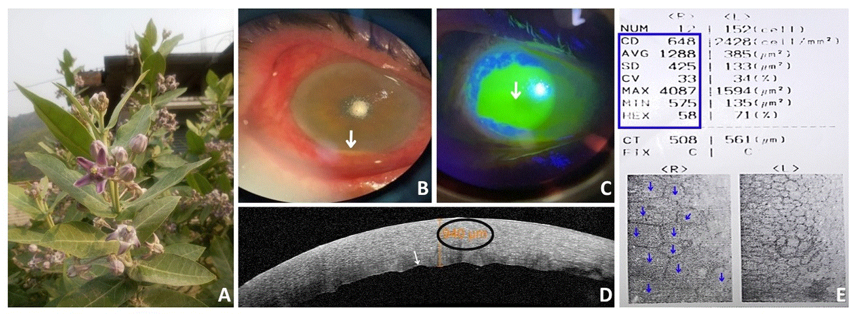
(A) Photograph of the Calotropis gigantea plant. (B) Slit-lamp photograph showing diffuse bulbar and circumciliary congestion, diffuse corneal edema with haze and a non-mobile inferior layered hypopyon (white arrow), and (C) fluorescein staining of large central corneal epithelial defect observed in cobalt blue light (white arrow). (D) Anterior segment optical coherence tomography (AS-OCT) showing corneal thickness of approximately 940 μm (black circle) with irregular undulating surface of descemet endothelial complex (white arrow). (E) Specular microscopy of the affected right eye showing reduced endothelial cell density (CD) (648 cells/mm2) (blue box) with marked polymegathism and pleomorphism (blue arrows).
Microbiological evaluation of a conjunctival swab, including Gram stain and potassium hydroxide (KOH) mount, revealed no bacterial or fungal organisms. Immediate copious irrigation of the ocular surface with normal saline was performed. Based on the history of Calotropis gigantea latex exposure, characteristic clinical findings, and the absence of organisms on initial microbiological evaluation, a diagnosis of Calotropis gigantea latex induced chemical ocular injury (RE toxic keratouveitis) was made. The patient was treated with topical moxifloxacin 0.5% 4 times a day, homatropine 2% 2 times a day, betaxolol 0.5% 2 times a day, sodium chloride 5% 4 times a day, carboxymethylcellulose 0.5%, ciprofloxacin 0.3% ophthalmic ointment, and fluorometholone 0.1% 4 times a day.
Over subsequent follow-up, the IOP decreased to 16 mmHg in both eyes. The corneal epithelial defect healed completely with gradual resolution of stromal edema, Descemet membrane folds, and anterior chamber inflammation, resulting in recovery of BCVA to 6/6 by 4 weeks. At the final follow-up, 1 year after the initial injury, the cornea remained clear with maintained BCVA of 6/6. However, specular microscopy demonstrated persistent severe endothelial cell loss, with an endothelial cell density (CD) of 648 cells/mm2 in the affected right eye ( Figure 1E) Marked polymegathism and pleomorphism indicated permanent toxic endothelial damage despite excellent anatomical recovery and good visual outcome ( Figure 1E).
Ocular exposure to Calotropis latex produces both direct cytotoxic and inflammatory damage to the cornea. Cardiac glycosides and proteolytic enzymes present in the latex disrupt epithelial integrity and impair endothelial pump function, resulting in corneal edema, Descemet membrane folds, keratouveitis, and transient visual loss.3–7 The clinical findings in our patient were consistent with previous reports describing conjunctival congestion, epithelial defects, stromal edema, and anterior chamber inflammation following Calotropis exposure.3–11
Multimodal ocular imaging provided objective documentation of both the acute injury and long-term sequelae. AS-OCT demonstrated marked corneal thickening (approximately 940 μm) with diffuse stromal hyperreflectivity, correlating with severe corneal edema observed clinically. This non-contact imaging modality complements slit-lamp examination by enabling quantitative assessment of corneal edema and monitoring structural recovery during follow-up.
The most significant finding was the persistent endothelial damage detected by specular microscopy one year after injury. Despite restoration of normal corneal clarity, central corneal thickness, and BCVA of 6/6, endothelial cell density remained markedly reduced (648 cells/mm2), with pronounced polymegathism and pleomorphism. Because human corneal endothelial cells have minimal regenerative capacity, injury is compensated by enlargement and morphological alteration of surviving cells rather than cellular regeneration.12 Specular microscopy is therefore indispensable for identifying subclinical endothelial dysfunction that may not be evident on routine clinical examination.13
Previous reports have primarily emphasized favorable visual recovery after Calotropis-induced ocular toxicity,3–11 whereas objective long-term endothelial assessment has rarely been documented. Our findings suggest that recovery of vision and corneal transparency does not necessarily indicate restoration of endothelial health. Patients with severe endothelial loss may remain at risk of future corneal decompensation, particularly following aging or intraocular surgery, highlighting the value of long-term follow-up with specular microscopy. In conclusion, Calotropis gigantea latex can cause significant corneal toxicity with permanent endothelial injury despite complete clinical and visual recovery. Multimodal ocular imaging was valuable for documenting both acute structural changes and persistent endothelial damage that was not clinically apparent. AS-OCT objectively quantified corneal edema, while specular microscopy revealed irreversible endothelial cell loss with polymegathism and pleomorphism. Long-term endothelial assessment should be considered in patients with moderate-to-severe Calotropis-induced ocular injuries to identify subclinical damage and guide prognosis.
Ethical approval was not required for this case report in accordance with the policies of the authors’ institution, as it describes a single anonymised patient managed with standard clinical care and involves no experimental intervention.
Written informed consent for publication was obtained from the patient.
No data are associated with this article.
The author(s) declared that no grants were involved in supporting this work.
© 2026 Pandey S et al. This is an open access article distributed under the terms of the Creative Commons Attribution License, which permits unrestricted use, distribution, and reproduction in any medium, provided the original work is properly cited.
Current Reviewer Status:
AWAITING PEER REVIEW
AWAITING PEER REVIEW
?
Key to Reviewer Statuses VIEW HIDE
ApprovedThe paper is scientifically sound in its current form and only minor, if any, improvements are suggested
Approved with reservations A number of small changes, sometimes more significant revisions are required to address specific details and improve the papers academic merit.
Not approvedFundamental flaws in the paper seriously undermine the findings and conclusions
Open Peer Review
AWAITING PEER REVIEW
Sign up for content alerts
Alongside their report, reviewers assign a status to the article:
Approved - the paper is scientifically sound in its current form and only minor, if any, improvements are suggested
Approved with reservations - A number of small changes, sometimes more significant revisions are required to address specific details and improve the papers academic merit.
Not approved - fundamental flaws in the paper seriously undermine the findings and conclusions