Просмотр новости
Найдите то, что Вас интересует
 Спутники
 РН
РН SpaceX
 Роскосмос
 NASA
NASA
СпутникиРНSpaceXРоскосмосNASAPurpose Hydrocephalus is a complex pediatric neurologic disorder characterized by abnormal cerebrospinal fluid (CSF) flow and ventricular dilation. It poses significant challenges in clinical management and often leads to severe morbidities despite treatment. This study explores the correlation between pre-shunt seizures in hydrocephalic children and inflammatory mediators and electrolyte imbalances in the CSF. Methods An analytical correlational study design was employed, utilizing a comprehensive array of clinical data, radiological databases, and cerebrospinal fluid (CSF) samples obtained from pediatric patients at seven neurosurgical centers. The study population consisted of children with hydrocephalus, both with and without a history of seizures. Sample selection followed strict inclusion and exclusion criteria. Laboratory examinations included a comprehensive CSF analysis for Na+, K+, Ca2+, Mg2+, IL-1β, IL-6, and TNF-α levels. Hydrocephalus imaging was based on established radiographic criteria, with particular focus on periventricular hypodensity. The analytical process explored the associations between inflammatory markers, electrolyte imbalances, periventricular hypodensity, and the occurrence of seizures. Result While most electrolytes showed no association with seizure activity, Mg2+ levels stood out as a notable exception. Specifically, higher Mg2+ levels were significantly correlated with seizure incidence, with a mean of 1.8 mEq/L (SD ± 0.4) and a p-value of 0.03. A significant difference in Ca2+ levels was also observed between hydrocephalus patients with marked periventricular hypodensity in the seizure and non-seizure groups. This finding was interesting because most studies have reported that hydrocephalic children with lower, not higher, electrolyte levels are more prone to seizures. Conclusion This study reveals significant associations between some electrolytes and seizure activity in pediatric hydrocephalus. Notably, Mg2+ levels were correlated with seizure incidence, suggesting a key role in seizure pathophysiology. Additionally, the observed differences in Ca2+ levels, particularly in patients with periventricular hypodensity, point to a potential connection between Ca2+ dysregulation, brain tissue alterations, and seizure susceptibility.

Sobana M, Gamayani U, Islam AA et al. A Comprehensive Analysis of Sodium, Potassium, Calcium, Magnesium,IL-1β, IL-6, and TNF-α, and Periventricular Hypodensity as Contributing Factors to Pre-Shunt Seizures in Hydrocephalic Children [version 1; peer review: 1 not approved]. F1000Research 2026, 15:1165 (https://doi.org/10.12688/f1000research.171165.1)
Research Article
[version 1; peer review: 1 not approved]
https://orcid.org/0000-0002-5208-9233
1, Uni Gamayanihttps://orcid.org/0000-0003-4707-8908
2, Andi Asadul Islam3, [...] Agnes Rengga Indrati4, Danny Halim5, Cut Vanessa Rachmadian1, Timotius Wira Yudha1, Rafela Agatha Christy3, Anggiat Silaen6, Meviraf Benny Tanio7, Priandana Adya Eka Saputra8, Elvira Sutanto9, Festus Andrianto Susilo10, Tri Hanggono Achmad11https://orcid.org/0000-0002-5208-9233
1, Uni Gamayanihttps://orcid.org/0000-0003-4707-8908
2, [...] Andi Asadul Islam3, Agnes Rengga Indrati4, Danny Halim5, Cut Vanessa Rachmadian1, Timotius Wira Yudha1, Rafela Agatha Christy3, Anggiat Silaen6, Meviraf Benny Tanio7, Priandana Adya Eka Saputra8, Elvira Sutanto9, Festus Andrianto Susilo10, Tri Hanggono Achmad111 Neurosurgery, Universitas Padjadjaran - Faculty of Medicine, Bandung, West Java, 40161, Indonesia
2 Neurology, Universitas Padjadjaran- Faculty of Medicine, Bandung, West Java, 40161, Indonesia
3 Neurosurgery, Hasanuddin University School of Medicine, Makassar, South Sulawesi, Indonesia
4 Clinical Pathology, Universitas Padjadjaran - Faculty of Medicine, Bandung, West Java, 40161, Indonesia
5 Neurosurgery, Siloam Hospitals Lippo Village, Tangerang, Banten, Indonesia
6 Neurosurgery, Hermina Hospital Pasteur, Bandung, West Java, Indonesia
7 Neurosurgery, Sayang Regional Hospital, Cianjur, West Java, Indonesia
8 Neurosurgery, Gunung Jati Hospital, Cirebon, West Java, Indonesia
9 Neurosurgery, Bandung Kiwari Regional Hospital, Bandung, West Java, Indonesia
10 Neurosurgery, Mitra Plumbon Hospital, Cirebon, West Java, Indonesia
11 Basic Medical Sciences, Universitas Padjadjaran- Faculty of Medicine, Bandung, West Java, 40161, Indonesia
Mirna Sobana
Roles: Conceptualization, Resources, Supervision
Uni Gamayani
Roles: Data Curation, Formal Analysis, Resources
Andi Asadul Islam
Roles: Investigation, Methodology, Writing – Review & Editing
Agnes Rengga Indrati
Roles: Formal Analysis, Validation, Visualization
Danny Halim
Roles: Data Curation, Writing – Original Draft Preparation
Cut Vanessa Rachmadian
Roles: Data Curation, Writing – Original Draft Preparation
Timotius Wira Yudha
Roles: Data Curation, Software, Writing – Review & Editing
Rafela Agatha Christy
Roles: Data Curation, Writing – Review & Editing
Anggiat Silaen
Roles: Data Curation, Writing – Original Draft Preparation
Meviraf Benny Tanio
Roles: Data Curation, Writing – Original Draft Preparation
Priandana Adya Eka Saputra
Roles: Data Curation, Writing – Original Draft Preparation
Elvira Sutanto
Roles: Data Curation, Writing – Original Draft Preparation
Festus Andrianto Susilo
Roles: Data Curation, Writing – Original Draft Preparation
Tri Hanggono Achmad
Roles: Conceptualization, Project Administration, Supervision, Validation, Visualization, Writing – Review & Editing
OPEN PEER REVIEW
REVIEWER STATUS
Hydrocephalus is a complex pediatric neurologic disorder characterized by abnormal cerebrospinal fluid (CSF) flow and ventricular dilation. It poses significant challenges in clinical management and often leads to severe morbidities despite treatment. This study explores the correlation between pre-shunt seizures in hydrocephalic children and inflammatory mediators and electrolyte imbalances in the CSF.
MethodsAn analytical correlational study design was employed, utilizing a comprehensive array of clinical data, radiological databases, and cerebrospinal fluid (CSF) samples obtained from pediatric patients at seven neurosurgical centers. The study population consisted of children with hydrocephalus, both with and without a history of seizures. Sample selection followed strict inclusion and exclusion criteria. Laboratory examinations included a comprehensive CSF analysis for Na+, K+, Ca2+, Mg2+, IL-1β, IL-6, and TNF-α levels. Hydrocephalus imaging was based on established radiographic criteria, with particular focus on periventricular hypodensity. The analytical process explored the associations between inflammatory markers, electrolyte imbalances, periventricular hypodensity, and the occurrence of seizures.
ResultWhile most electrolytes showed no association with seizure activity, Mg2+ levels stood out as a notable exception. Specifically, higher Mg2+ levels were significantly correlated with seizure incidence, with a mean of 1.8 mEq/L (SD ± 0.4) and a p-value of 0.03. A significant difference in Ca2+ levels was also observed between hydrocephalus patients with marked periventricular hypodensity in the seizure and non-seizure groups. This finding was interesting because most studies have reported that hydrocephalic children with lower, not higher, electrolyte levels are more prone to seizures.
ConclusionThis study reveals significant associations between some electrolytes and seizure activity in pediatric hydrocephalus. Notably, Mg2+ levels were correlated with seizure incidence, suggesting a key role in seizure pathophysiology. Additionally, the observed differences in Ca2+ levels, particularly in patients with periventricular hypodensity, point to a potential connection between Ca2+ dysregulation, brain tissue alterations, and seizure susceptibility.
Hydrocephalus, Seizures, Cerebrospinal fluid, Inflammation, Electrolytes, Periventricular Hypodensity
Corresponding author: Mirna Sobana Competing interests: No competing interests were disclosed.
Grant information: The author(s) declared that no grants were involved in supporting this work.
Copyright: © 2026 Sobana M et al. This is an open access article distributed under the terms of the Creative Commons Attribution License, which permits unrestricted use, distribution, and reproduction in any medium, provided the original work is properly cited. How to cite: Sobana M, Gamayani U, Islam AA et al. A Comprehensive Analysis of Sodium, Potassium, Calcium, Magnesium,IL-1β, IL-6, and TNF-α, and Periventricular Hypodensity as Contributing Factors to Pre-Shunt Seizures in Hydrocephalic Children [version 1; peer review: 1 not approved]. F1000Research 2026, 15:1165 (https://doi.org/10.12688/f1000research.171165.1) First published: 15 Jul 2026, 15:1165 (https://doi.org/10.12688/f1000research.171165.1) Latest published: 15 Jul 2026, 15:1165 (https://doi.org/10.12688/f1000research.171165.1)
Hydrocephalus is a neurological condition, either congenital or acquired, caused by impaired CSF flow, leading to ventricular dilation.1–3 While CSF diversion is the main treatment, hydrocephalic children remain at risk for morbidities like cerebral palsy, seizures, and sensory impairments. A meta-analysis shows a 15.75 times higher risk of seizures in shunted hydrocephalic children, with some experiencing seizures before shunt placement, suggesting a link between pre-shunt seizures and hydrocephalus.4–8
CSF diversion provides a definitive solution, yet pre-shunt seizures suggest an underlying pathogenesis in hydrocephalus. In pre-shunt hydrocephalus, increased intracranial pressure (ICP) results from the accumulation of cerebrospinal fluid, while decreased cerebral blood flow (CBF) and cerebral perfusion pressure (CPP) occur due to the compression of brain tissues and blood vessels, leading to reduced oxygen and nutrient delivery to the brain. This irritates the brain cells, which in turn help trigger seizures.9 Our study explored the link between periventricular hypodensity and pre-shunt seizures, finding a strong correlation. This hypodensity, indicating ependymal damage, CSF leakage, and periventricular hypoplasia, is linked to ventricular dilation and inflammation. While the normal ependymal lining forms a CSF-brain barrier, inflammation may cause the barrier to fail. One such case in an infant demonstrated a white matter injury after inflammation caused by COVID-19 infection.
We propose that CNS inflammation, marked by elevated IL-1β, and IL-6, and TNF-α is associated with seizures, as are electrolyte imbalances in Na+, K+, Ca2+, and Mg2+. These markers were chosen due to the availability of assay kits in our centre. This study aims to investigate the role of these electrolyte imbalances, inflammatory mediators, and periventricular hypodensity in the development of seizures in children with hydrocephalus, uncovering the pathogenesis of pre-shunt seizures in this population.10–12 The findings may identify these factors as potential biomarkers for pre-shunt seizures, providing insights for improved clinical management.
This research employs an analytical correlational study design. The algorithms of this study are as follows: Pediatric patients under 18 years old admitted to the Department of Neurosurgery at the following hospitals: Hasan Sadikin General Hospital (Bandung), Wahidin Sudirohusodo General Hospital (Makassar), Hermina Hospital Pasteur (Bandung), Sayang Hospital (Cianjur), Siloam Putera Bahagia Hospital (Cirebon), Bandung Kiwari Hospital (Bandung), Siloam Hospital Lippo Village, and Mitra Plumbon Hospital (Cirebon); underwent comprehensive clinical examination. Based on the findings, patients suspected of having hydrocephalus were classified into two groups: those with seizures and those without seizures. Patients were excluded if they: lacked patient or family consent; had a seizure history unrelated to hydrocephalus; had a family history of epilepsy; or were undergoing treatment with anti-seizure or anti-inflammatory medication. Written informed consent was obtained from the parents or legal guardians of all participating children. Because several children were under the legal age of 18, verbal assent was not required. All procedures were reviewed and approved by the Research Ethics Committee of Padjadjaran University (Approval No. 2204050469).
Patients then underwent radiological examinations to confirm the diagnosis of hydrocephalus. The diagnostic criteria for hydrocephalus include indicators such as widened temporal horns, an Evans' ratio exceeding 30%, an increased frontal horn-to-internal-diameter ratio (>50%), dilation of the frontal horns of the lateral ventricles, periventricular hypodensity, and upward bowing of the corpus callosum. Children diagnosed with hydrocephalus who were planned for and had provided consent for ventriculoperitoneal (VP) shunt placement were included in this study.
Cerebrospinal fluid (CSF) were isolated intraoperatively during the initial ventricular puncture. A sample volume of 5 mL were collected for analysis. Laboratory procedures include measurement of electrolyte levels (Na+, K+, Ca2+, Mg2+), and cytokine assays (IL-1β, IL-6, TNF-α) were conducted in accordance with standardized protocols. CSF samples for transport were collected in sterile polypropylene tubes, aliquoted to minimize freeze-thaw cycles, and immediately refrigerated at 2-8°C. For transport exceeding 24 hours or requiring long distances, samples were frozen at -20°C. Specialized transport containers with temperature monitoring capabilities and sufficient dry ice or coolant packs were used to maintain the desired temperature range throughout the journey.13–15
The sample collection for this study employs a stratified sampling method, targeting hydrocephalic children who meet specific inclusion criteria from March 1st 2022, to February 28th, 2024, where patients are grouped according to their seizure status (seizure vs no seizure). The serum samples were taken upon the child’s initial admission to the hospital.
The dependent variables are the concentrations of Na+, K+, Ca2+, Mg2+, IL-1β, IL-6, and TNF-α in both serum and CSF. The independent variables include the diagnosis of hydrocephalus, periventricular hypodensity, and presence of pre-shunt seizures. The explained variables were operationalized with measuring instruments to obtain the expected results for the study. The operational definitions of the variables are: Seizure, determined based on patient history as recorded in the medical records, indicating whether seizures are present or not; Hydrocephalus, diagnosis based on imaging results confirming the presence of hydrocephalus according to established criteria; Electrolytes, such as Na+ (Na+), K+ (K+), Ca2+ (Ca2+), Mg2+ (Mg2+), with concentrations measured in both serum and cerebrospinal fluid (CSF) samples using ion-selective electrolyte assays, reported in millimoles per liter (mmol/L); and Inflammatory mediators, such as IL-1β (Interleukin-1 beta), IL-6 (Interleukin-6), and TNF-α (Tumor Necrosis Factor-alpha), with levels detected in serum and CSF samples using cytometric bead array analysis, reported in picograms per milliliter (pg/mL).
Data processing involves the entry, cleaning, and editing of raw data to ensure reliability before subsequent analysis. Descriptive statistics and appropriate statistical tests (t-test, Wilcoxon signed-rank test, chi-square goodness of fit) were employed for data analysis. Following data collection, we first outlined the general characteristics of the subjects. Then independent t-tests were performed on electrolyte and inflammatory markers, among the seizure and non-seizure groups, and significant associations were noted. In addition, we also tested whether periventricular hypodensity was associated with pre-shunt seizures, via chi-squared test. This triggered the curiosity for assessing the independent t-tests on the earlier parameters, but only on subjects with periventricular hypodensity (as exemplified in Figure 1). Finally, some explorations were also performed using correlation tests on significant electrolytes versus inflammatory markers.
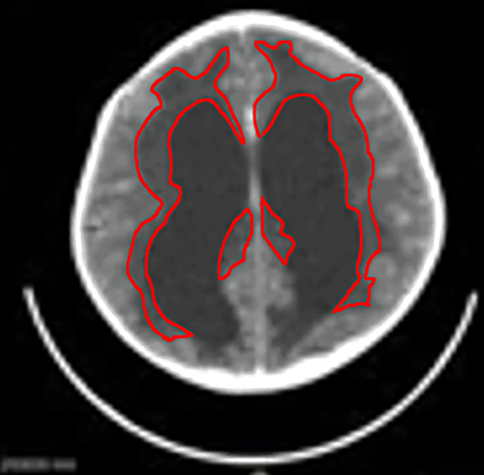
In the statistical analysis of electrolytes (Na+, K+, Ca2+, Mg2+) and inflammatory mediators in cerebrospinal fluid (CSF) of hydrocephalic children with pre-shunt seizures, the data revealed no significant associations between these electrolytes and the incidence of seizures, with the notable exception of Mg2+, and in the periventricular hypodensity group particularly, Ca2+. The general characteristics of the patients are shown in Table 1.
We collected a total number of 44 participants, ensuring robust data for analysis. Out of these samples, 18 had seizures and 26 did not experience them. The dataset reveals a notable trend among children with hydrocephalus, indicating that a significant proportion (41%, n = 18) experienced seizures. The majority of the participants were under one year old (59%, n = 26), suggesting that younger children may be more susceptible to seizure activity. Additionally, the presence of periventricular hypodensity was observed in over half of the cases (55%, n = 24), which may correlate with the incidence of seizures. Indeed, in the seizure group, 16 out of 18 (89%) had periventricular hypodensity, while only two (11%) did not have. Whereas in the no seizure group, periventricular hypodensity was found in only eight out of 26 (31%), compared to 18 (69%) who did not have.
The comparison of inflammatory mediators between seizure and non-seizure populations, as seen in Table 2, reveals that the seizure group exhibited mean levels of IL-1β at 23.18 pg/mL, IL-6 at 49.87 pg/mL, and TNF-α at 101.91 pg/mL. In contrast, the non-seizure group had mean levels of IL-1β at 13.06 pg/mL, IL-6 at 28.10 pg/mL, and TNF-α at 40.06 pg/mL. The p-values for IL-1β, IL-6, and TNF-α were 0.260, 0.669, and 0.224, respectively, indicating that there were no statistically significant differences in the levels of these inflammatory mediators between the two groups.
The analysis of electrolyte levels between the seizure and non-seizure populations reveals that Na+ (Na+) and K+ (K+) levels did not exhibit significant differences, with means of 130.4 mEq/L and 2.63 mEq/L in the seizure group compared to 133.1 mEq/L and 2.36 mEq/L in the non-seizure group, respectively. However, Ca2+ (Ca2+) levels approached significance, showing a mean of 1.81 mEq/L in the seizure group versus 1.49 mEq/L in the non-seizure group (p=0.079). Notably, Mg2+ (Mg2+) levels were significantly higher in the seizure group at 1.98 mEq/L compared to 1.57 mEq/L in the non-seizure group, with a p-value of 0.049. This suggests a potential link between elevated Mg2+ levels and seizure activity in this population.
As by distinguishing samples by their periventricular hypodensity status, it revealed that none of the inflammatory markers or the electrolytes appeared to be statistically significant.
The analysis of children identified with periventricular hypodensity on CT-scan indicates that Na+ and K+ levels did not show significant differences between the seizure group and the non-seizure group, which was visible in Table 3. We found that means of 131.2 mEq/L and 2.76 mEq/L in the seizure group compared to 123.8 mEq/L and 2.55 mEq/L in the non-seizure group, respectively. However, Ca2+ levels were significantly higher in the seizure group, averaging 3.79 mEq/L, compared to 2.25 mEq/L in the non-seizure group, with a p-value of 0.001, indicating strong statistical significance. Other inflammatory markers, including TNF-alpha, IL-1ß, and IL-6, did not demonstrate significant differences between the two groups, suggesting that while Ca2+ levels may be associated with seizure activity, the inflammatory markers assessed do not appear to differ significantly in this population.
Table 4 presents the statistical analysis of the relationship between periventricular hypodensity and seizures. Among the subjects, 18 individuals without seizures also did not exhibit periventricular hypodensity, while only 2 individuals without seizures showed the presence of hypodensity. Conversely, 8 individuals with periventricular hypodensity did not experience seizures, whereas 16 individuals with seizures also presented with hypodensity. The results indicate a significant association between the presence of periventricular hypodensity and the occurrence of seizures, with a p-value of less than 0.001.
We also explored the potential electrolyte differences on samples with periventricular hypodensity and those without.
As Mg2+ turned out to be statistically significant factor, a correlation analysis was performed between Mg2+ and the three inflammatory markers. Table 5 presents the results of the correlation test between Mg2+ levels and various inflammatory markers. The correlation between Mg2+ and TNF-alpha is -0.91, with a p-value of 0.573, indicating no significant relationship. Similarly, the correlation with IL-1ß is -0.142 (p-value 0.376) and with IL-6 is -0.056 (p-value 0.728), both suggesting a lack of significant correlation between Mg2+ levels and these inflammatory markers. These findings imply that Mg2+ levels do not appear to influence the inflammatory response in this population.
Additionally with Ca2+ in the periventricular hypodensity group, we explored the correlation between this ion and the three inflammatory markers, which are presented in Table 6. Not only all inflammatory markers turned out not to be significant (with p-values of 0.431, 0.706, and 0.707 respectively for TNF-alpha, IL-1ß, and IL-6); their correlations were also very low (0.169, -0.081, and -0.081 for each). This suggests that Ca2+ levels also did not influence the inflammatory response in the periventricular hypodensity group.
The dataset provides a comprehensive analysis of the role of electrolytes in the occurrence of seizures among children with hydrocephalus. Notably, Table 2 highlights a significant difference in Mg2+ levels between seizure and non-seizure groups in the total cohort (p = 0.049), suggesting that Mg2+ may play a critical role in seizure activity. Conversely, Ca2+ levels did not show a significant difference in this cohort, indicating that while Mg2+ may be a key player, its role may be less pronounced in the periventricular hypodensity cohort. Mg2+ is an essential cofactor in numerous enzymatic reactions and is crucial for maintaining neuronal function. It acts as a natural Ca2+ antagonist, regulating Ca2+ influx into neurons, which is vital for neurotransmitter release and neuronal excitability. Although traditionally hypomagnesemia is thought as a major contributor to seizures, a higher level of this ion has been documented with seizures, especially in conjunction with hypocalcemia.16
In contrast, Table 3, which focuses on the periventricular hypodensity subset, reveals that Ca2+ levels were significantly different (p = 0.001), while Mg2+ levels did not show a significant difference. This finding suggests that in the context of periventricular hypodensity, Ca2+ may play a more critical role in seizure susceptibility than Mg2+. The presence of periventricular hypodensity may indicate underlying white matter injury, which can disrupt normal neuronal circuitry and exacerbate the effects of low Mg2+ levels. This disruption can create a feedback loop, where low Mg2+ levels contribute to increased seizure susceptibility, and the resulting seizures may further impact Mg2+ homeostasis.17
Furthermore, the study by Castilla-Guerra et al.2 emphasized that electrolyte disturbances can trigger seizures, reinforcing the need to monitor Mg2+ and Ca2+ levels in hydrocephalic patients. Recent studies have further elucidated the importance of Na+ and Ca2+ imbalances in pediatric populations. For instance, a study by Meisler et al. (2005) demonstrated that Na+ channel dysfunction can lead to increased seizure susceptibility in children, reinforcing the need to monitor Na+ levels in hydrocephalic patients. Additionally, Ca2+'s role in neurotransmitter release and neuronal excitability has been highlighted by Zhou et al., who found that Ca2+ dysregulation is associated with seizure activity in children with neurological disorders.18,19 Despite no significant associations were found with Na+ in this study, we still believe it plays a critical role in seizures and can be replicated in further studies.
The lack of significant differences in K+ levels indicates that while these electrolytes are essential for neuronal excitability, their roles may be altered in the context of hydrocephalus. Kaplan et al. also noted the importance of Na+ channels in epilepsy, suggesting that the unique pathophysiological mechanisms in hydrocephalic children warrant further investigation into the specific roles of these electrolytes in seizure susceptibility. Overall, the interplay between Mg2+ and Ca2+ levels, periventricular hypodensity, and seizure activity highlights the need for a multifaceted approach to understanding and managing seizures in children with hydrocephalus, particularly in light of the potential neuroprotective effects of Mg2+ and its role in maintaining neuronal stability.18,20
Inflammation's role in seizure activity among children with hydrocephalus is explored in Table 4, which presents inflammatory mediators in both seizure and non-seizure groups. Although no significant differences were observed, the elevated levels of TNF-α and IL-6 in the seizure group warranted further investigation as an inflammatory component in seizure pathogenesis. This is consistent with findings by Webster et al., who highlighted the significance of inflammation in the development of epilepsy. Recent research by Choi et al. has shown that elevated levels of pro-inflammatory cytokines, including IL-1β and IL-6, are associated with increased seizure frequency in pediatric populations, indicating that inflammation may contribute to seizure susceptibility. However, the lack of significant correlations between inflammatory markers and seizure activity in our study indicates that inflammation may not play a primary role in this specific population. Future research should investigate the nuances of inflammatory processes in hydrocephalus and their potential contributions to seizure susceptibility, particularly in light of emerging evidence linking chronic inflammation to neurodevelopmental disorders.4,21
The association between periventricular hypodensity and seizures is strongly supported by Table 5, which shows a significant correlation (p < 0.001). This finding underscores the importance of structural brain changes in understanding seizure risk among hydrocephalic children. Sood et al. (2005) noted that long-term shunt treatment for hydrocephalus can lead to periventricular rigidity, which may further complicate seizure susceptibility. Recent studies, have demonstrated that periventricular white matter injury is associated with an increased risk of seizures in children, suggesting that the presence of periventricular hypodensity may indicate underlying white matter injury, disrupting normal neuronal circuitry and increasing the likelihood of seizures. Kulkarni et al. also explored structural interventions in hydrocephalus treatment, emphasizing the need for comprehensive neuroimaging assessments to better understand the relationship between structural abnormalities and seizure activity. The implications of these findings highlight the necessity for early identification and management of periventricular hypodensity to mitigate seizure risk in this vulnerable population.22–25
The interplay between Mg2+ levels, inflammatory markers, and periventricular hypodensity presents a complex relationship that warrants further exploration. Table 6 reveals weak correlations between Mg2+ and inflammatory markers, suggesting that while Mg2+ levels are altered in seizure patients, their direct relationship with inflammation markers like TNF-α and IL-6 is minimal. This aligns with Steensberg et al. (2006), who noted the complex interplay of inflammation in neurological conditions. Recent findings by Nielsen et al. (2018) indicate that low Mg2+ levels may exacerbate inflammatory responses, potentially creating a feedback loop that increases seizure susceptibility. Additionally, the presence of periventricular hypodensity may exacerbate the effects of low Mg2+ levels, creating a feedback loop that increases seizure susceptibility. Understanding this three-way relationship is crucial for developing comprehensive management strategies that address the multifactorial nature of seizures in hydrocephalic children. Future research should aim to elucidate the specific pathways through which these variables interact, as this knowledge could inform targeted therapeutic interventions that optimize patient care.21,24,26
This study could have benefited more if the degrees of parameters could be differentiated semi-quantitatively. For example, hydrocephalus could be further classified as mild vs marked; periventricular hypodensity as mild or intense.
At this moment, these distinctions could not be fulfilled, due to the heterogeneity of radiographic devices used during the collection (multi-centre) as with the imaging.
Secondly, owing to the limitations of the software at the moment, multivariate analysis could have been employed in future studies.
Some electrolytes, like Mg2+ in general, and Ca2+ in the periventricular hypodensity group, were associated with seizures in children with hydrocephalus. Along with periventricular hypodensity, they emerged as critical factors in the pathogenesis of these seizures. Future research could improve the methodology for electrolyte/marker sample collection and analysis, with a focus on detailed imaging studies and inflammatory markers. This would further elucidate these relationships and help develop targeted interventions for hydrocephalic children at risk for seizures. This shift in research focus not only broadens the understanding of hydrocephalus-related seizures but also opens avenues for improved clinical management strategies.
This study has received ethical approval from the Research Ethics Committee of the Padjadjaran University, with registration number 2204050469.
OSF: Supplementary Files to “A Comprehensive Analysis of Sodium, Potassium, Calcium, Magnesium, IL-1β, IL-6, TNF-α, and Periventricular Hypodensity as Contributing Factors to Pre-Shunt Seizures in Hydrocephalic Children”. https://doi.org/10.17605/OSF.IO/V27FE.27
The project contains the following underlying data:
- SPSS Data hydrocephalus seizure English.sav, contains the raw data in SPSS format.
- IC.pdf, contains the form given to patients’ parents for obtaining consent.
- English Translation – Informed Consent (hydrocephalus Study).pdf, contains the English translation for the consent forms
Data are available under the terms of the Creative Commons Attribution 4.0 International license (CC-BY 4.0).
The author(s) declared that no grants were involved in supporting this work.
© 2026 Sobana M et al. This is an open access article distributed under the terms of the Creative Commons Attribution License, which permits unrestricted use, distribution, and reproduction in any medium, provided the original work is properly cited.
Current Reviewer Status: ?
Key to Reviewer Statuses VIEW HIDE
ApprovedThe paper is scientifically sound in its current form and only minor, if any, improvements are suggested
Approved with reservations A number of small changes, sometimes more significant revisions are required to address specific details and improve the papers academic merit.
Not approvedFundamental flaws in the paper seriously undermine the findings and conclusions
Version 1
VERSION 1
PUBLISHED 15 Jul 2026
Reviewer Report 05 Aug 2026
Casper Schwartz Riedel, Copenhagen University, Copenhagen, Denmark
Not Approved
VIEWS 0
Is the work clearly and accurately presented and does it cite the current literature?
Partly
Is the study design appropriate and is the work technically sound?
Partly
Are sufficient details of methods and analysis provided to allow replication by others?
No
If applicable, is the statistical analysis and its interpretation appropriate?
Partly
Are all the source data underlying the results available to ensure full reproducibility?
Yes
Are the conclusions drawn adequately supported by the results?
Partly
Competing Interests: No competing interests were disclosed.
Reviewer Expertise: Neurosurgery, neuroradiology, hydrocephalus, sleep apnea and intracranial pressure.
CloseOpen Peer Review
Alongside their report, reviewers assign a status to the article:
| Invited Reviewers | |
|---|---|
| 1 | |
| Version 1 15 Jul 26 | read |
Sign up for content alerts
Alongside their report, reviewers assign a status to the article:
Approved - the paper is scientifically sound in its current form and only minor, if any, improvements are suggested
Approved with reservations - A number of small changes, sometimes more significant revisions are required to address specific details and improve the papers academic merit.
Not approved - fundamental flaws in the paper seriously undermine the findings and conclusions