Просмотр новости
Найдите то, что Вас интересует
 Спутники
 РН
РН SpaceX
 Роскосмос
 NASA
NASA
СпутникиРНSpaceXРоскосмосNASAIntroduction Septic shock is a condition resulting from an uncontrolled immune response to infection, leading to multi-organ failure. Fluid resuscitation is the primary initial therapy, but an aggressive approach risks fluid overload and increased extravascular lung water (EVLW). To date, direct comparative evidence between fluid-based resuscitation strategies, vasopressors, or a combination of both on hemodynamic changes is limited. This study aims to evaluate the differences in hemodynamic responses of these three strategies using the Sus scrofa domestica animal model. Methods This experimental study, conducted at the Veterinary Teaching Hospital of IPB University from November 2024 to January 2025, comprised 15 male Sus scrofa domestica, aged 16–20 weeks, weighing 40–60 kg, plus three controls. Randomization was performed using Random.org with allocation concealment, and procedures were standardized to minimize confounding. Septic shock was induced using lipopolysaccharide (E. coli O111:B4) until a MAP of
Septic shock is characterized by an uncontrolled and excessive immune response to infection, which causes endothelial dysfunction, systemic vasodilation, increased vascular permeability, and impaired tissue perfusion leading to multi-organ failure.1,2 Septic shock remains a major issue as decades of research for a therapy that successfully address the host dysregulated, overshooting immune response is unavailable.3 Fluid resuscitation is a key component in the initial management of septic shock, however, an aggressive fluid resuscitation approach has received attention due to the potential for fluid overload, pulmonary edema, and increased extravascular lung water (EVLW).4,5
Understanding of the hemodynamic pathophysiology of septic shock has evolved regarding the role of systemic vasodilation and decreased systemic vascular resistance (SVR). This underlies the use of vasopressor as first-line therapy to maintain perfusion pressure by increasing vascular tone without significantly decreasing cardiac output.6 Meta-analysis showed that norepinephrine is effective in achieving target mean arterial pressure (MAP) and has a better safety profile than other vasopressors, although its impact on mortality is still variable.1
However, evidence regarding direct comparisons between fluid-based resuscitation strategies alone, vasopressors alone, or a combination of both on hemodynamic changes is still limited.7 We developed a Sus scrofa model as a sepsis animal model to study the hemodynamic differences between resuscitation using fluids alone, fluids with norepinephrine, and norepinephrine alone. We hypothesize that the combined fluid and norepinephrine strategy will achieve significantly different in hemodynamic response.
This experimental study was conducted from November 2024 to January 2025, at the Veterinary Teaching Hospital of the School of Veterinary Medicine and Biomedical Sciences, IPB University, Bogor, Indonesia. This research was approved by the animal ethics committee of the school of veterinary medicine and biomedicine, IPB University 266/KEH/SKE/2/2024. This research was conducted in accordance with the ARRIVE (Animal Research: Reporting of in vivo Experiments) guidelines.8,9
Animals were included if they met the following criteria: (1) male Sus scrofa domestica, (2) aged 16–20 weeks, (3) body weight 40–60 kg, and (4) developed septic shock during the experiment. Animals were excluded if they had been used in previous experiments or died from sepsis or septic shock prior to completion of data collection. The estimated sample size was determined using the resource equation10
E=N−T
10=(Nx3)−3
13=Nx3
N=13/3=4.34
Where:
E: Between 10–20.
N: Number of animals per treatment group multiplied by the number of treatment groups.
T: Number of treatments per group.
The sample size for this study was 4 Sus scrofa domestica calves per group. There were 3 groups in this study, so the total sample size required was 12 Sus scrofa domestica. The total sample size required was 15 Sus scrofa domestica in addition with 3 Sus scrofa domestica in control group.
Randomization in this study was conducted using the Random.org website by a single research assistant who was not directly involved in the intervention process or outcome assessment. The results of the group allocation were not known to the rest of the research team until the time of treatment administration (allocation concealment). To reduce potential confounding factors, all study procedures were standardized, including a consistent order of treatment administration, uniform measurement times for each subject, and randomized subject assignment. Participants and outcome assessors were blinded to group allocation. The intervention was prepared and administered by designated personnel who were not involved in data collection or analysis. Data analysts were also blinded to group assignments during statistical analysis.
Equipment used included a CHI-SON Ebit60 Vet® ultrasound machine (CHISON Medical Technologies Co., Ltd., China), a Mindray® MEC 1000 patient monitor, and a transpulmonary thermodilution catheter system (PiCCO, PULSION Medical Systems SE, Munich, Germany), along with standard surgical instruments, ventilator support, and monitoring devices.
Pharmacological agents included ketamine 10%, xylazine 2%, fentanyl, propofol 0.1%, isoflurane 100%, norepinephrine, crystalloid solution (Ringer-Lactate), povidone-iodine 10%, alcohol 70%, marbofloxacin (Marbocyl® 10%), Hematodin®, Bio-din®, ivermectin®, and endotoxin lipopolysaccharide (E. coli 0111:B4). ELISA kits were used for measurement of IFABP, NT-proBNP, Syndecan-1, NGAL, and cystatin-C.
The overall experimental setup and workflow are illustrated in Figure 1. Animals underwent a 10-day acclimatization period under controlled environmental conditions (temperature 18–26 °C; humidity >80%) with standard feeding twice daily and free access to water. Feeding was withheld for 6 hours prior to experimentation. During acclimatization, animals received prophylactic treatment consisting of marbofloxacin (1 mL/10 kg BW for 7 days), Hematodin® and Bio-din® (2 mL/50 kg BW on days 1, 3, and 7), ivermectin® (1 mL/50 kg BW subcutaneously), and fenbendazole (4 mL/50 kg BW orally).11,12
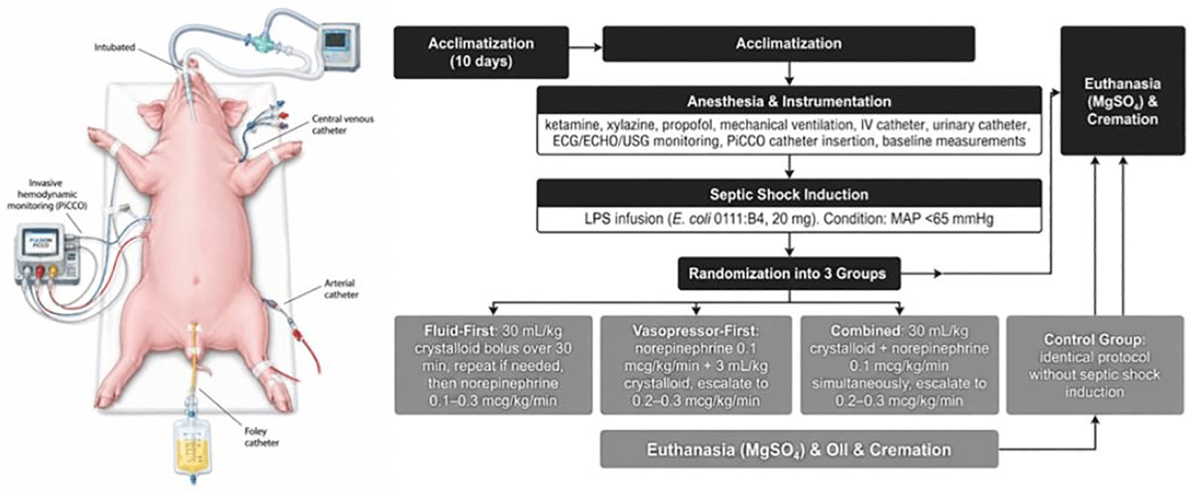
Animals were anesthetized, intubated, and mechanically ventilated, with placement of arterial and central venous catheters, as well as a PiCCO catheter for transpulmonary thermodilution monitoring. Septic shock was induced using lipopolysaccharide (E. coli O111:B4), followed by randomization into fluid-first, vasopressor-first, and combined resuscitation groups. Hemodynamic parameters and laboratory measurements were recorded according to the study protocol.
Anesthesia was induced using intramuscular ketamine (10 mg/kg BW) and xylazine (2 mg/kg BW), followed by endotracheal intubation after propofol administration (4–6 mg/kg BW). Animals were mechanically ventilated (tidal volume 8 mL/kg, PEEP 4 cmH₂O, FiO₂ 0.4), and anesthesia was maintained with isoflurane (2%). A 22G intravenous catheter was inserted into the auricular vein for fluid administration (0.9% NaCl at 3 mL/kg/hour). Urinary catheterization was performed. Hemodynamic monitoring included electrocardiography (ECG), echocardiography (ECHO), and ultrasonography (USG). After stabilization, a PiCCO catheter was inserted via the jugular vein. Baseline blood samples and hemodynamic measurements were obtained within 10–15 minutes after stabilization. Septic shock was induced using intravenous infusion of lipopolysaccharide (E. coli 0111:B4; 20 mg diluted in 20 mL sterile water). Septic shock was defined as a decrease in MAP to <65 mmHg.3,13
Following septic shock induction, animals were randomized into three resuscitation strategies. In the fluid-first group, resuscitation was initiated with a 30 mL/kg crystalloid bolus administered over 30 minutes and repeated if the target mean arterial pressure (MAP ≥65 mmHg) was not achieved; persistent hypotension was managed with norepinephrine titrated from 0.1 to 0.3 mcg/kg/min. In the vasopressor-first group, norepinephrine was initiated at 0.1 mcg/kg/min and combined with a 3 mL/kg crystalloid bolus, with dose escalation to 0.2–0.3 mcg/kg/min as needed to achieve the target MAP. In the combined group, animals received simultaneous administration of a 30 mL/kg crystalloid bolus and norepinephrine at 0.1 mcg/kg/min, followed by stepwise escalation of norepinephrine (0.2–0.3 mcg/kg/min) if the target MAP was not reached. Control groups followed identical protocols without induction of septic shock. At the conclusion of the experiment, animals were euthanized under anesthesia using magnesium sulfate (MgSO₄) and subsequently cremated. This method was selected because it provides rapid cardiac arrest in anesthetized animals.
The study outcomes were assessed based on: Achievement of the target MAP ≥65 mmHg. Primary outcome: Hemodynamics: MAP, CI, SVRI, SVI, GEDVI, ELWI. Secondary outcome: IFABP, lactate, Hb, Oxygen transport: DO₂ and CaO₂. Furthermore, treatment failure was defined as failure to achieve the target MAP after three protocol escalations. Samples that failed to reach the MAP target were still included in the study. The amount of fluid is determined based on: Bolus administered according to the group protocol (30 mL/kg or 3 mL/kg). Repeat bolus administration if the target MAP of ≥65 mmHg has not been achieved. There is no other specific method for measuring total fluid volume (e.g., cumulative fluid balance).
Animal welfare was prioritized in accordance with institutional ethical standards and the ARRIVE 2.0 guidelines. Clinical signs of pain, distress, or physiological deterioration were documented and assessed by the research team and attending veterinarian. To minimize pain and distress, all invasive procedures were performed under general anesthesia induced with ketamine and xylazine and maintained with propofol and isoflurane as described in the protocol.
Data were presented as mean ± standard deviation for normally distributed variables and median (minimum–maximum) for non-normally distributed variables. Normality testing was performed prior to analysis. Comparisons between two groups were conducted using Independent t-test or Mann–Whitney U test, while paired comparisons used Paired t-test or Wilcoxon test. Comparisons among three or more groups were analyzed using One-way ANOVA or Kruskal–Wallis test. Correlation analyses were performed using Pearson or Spearman tests as appropriate. Statistical significance was defined as p < 0.05. Statistical analyses were performed using IBM SPSS Statistics version 32 (IBM Corp., Armonk, NY, USA).
The current protocol was created to assess three resuscitation techniques: fluid-first, vasopressor-first, and combined fluid-vasopressor therapy in controlled porcine model of septic shock. Porcine are one of the most relevant large-animal models for sepsis research because their cardiovascular system, size, and blood flow responses closely resemble those of humans.14 Recent reviews highlight that porcine models of septic shock offer better insight compared to small-animal models. They also allow monitoring methods that closely mimic intensive care management in human patients.15 A key strength of this study is the use of invasive transpulmonary thermodilution monitoring (PiCCO). This allows detailed study of preload, cardiac performance, vascular resistance and fluid accumulation in the lungs.16 The study is also expected to contribute to the ongoing debate about initiation of vasopressor treatment. There is increasing evidence that earlier initiation of vasopressors can contribute to the reduction of the amount of intravenous fluids needed for hemodynamic stability. This may contribute to prevention of fluid overload and tissue swelling.17,18 However, adequate fluid resuscitation remains critical to optimize heart preload and to ensure proper reaction to vasopressor therapy.19,20 By comparing fluid-first, vasopressor-first and combined approaches under controlled conditions, this study may provide insight into the interaction between vascular tone, blood volume and oxygen delivery during septic shock.
Several limitations need to be noted. Even with careful standardization, biological differences among the animals can affect their responses to endotoxin exposure and resuscitation. Variations in initial blood volume might also impact the ability to reach target MAP and other blood flow outcomes.21 Endotoxin-induced septic shock mimics many hemodynamic features of human sepsis. However, it may not completely represent the complexity of real clinical infections where pathogen-host interactions occur.22
This study is expected to provide an essential experimental platform to evaluate hemodynamic responses in septic shock using a controlled Sus scrofa domestica model. By directly comparing fluid-first, vasopressor-first, and combined resuscitation strategies, this study will generate mechanistic insights into the interplay between intravascular volume, vascular tone, and tissue perfusion, particularly in achieving target mean arterial pressure (MAP).
The challenge in this study is the potential for uncontrolled differences in intravascular volume between subjects, which could influence the hemodynamic response to resuscitation, potentially confounding the achievement of MAP targets and changes in other parameters such as ELWI and DO₂. Furthermore, the relatively small sample size makes it likely that some clinically significant differences may not reach statistical significance.
The results of this study will be disseminated through publication in a peer-reviewed open-access journal and presentation at relevant scientific conferences. The findings are expected to contribute to the understanding of hemodynamic resuscitation strategies in septic shock and may inform future experimental and clinical research. Any data generated from this study will be made available in an appropriate public repository in accordance with journal policies.
No data are associated with this article. Open Science Framework.
The authors would like to thank the staff of the Veterinary Teaching Hospital, School of Veterinary Medicine and Biomedical Sciences, IPB University, Bogor, Indonesia, for their technical assistance in research process.