Просмотр новости
Найдите то, что Вас интересует
 Спутники
 РН
РН SpaceX
 Роскосмос
 NASA
NASA
СпутникиРНSpaceXРоскосмосNASABackground Tuberculosis (TB) remains a significant health issue in Indonesia, ranking second globally in 2024. Diagnosing intestinal tuberculosis (ITB) is challenging due to its symptoms mimicking other diseases and limited non-invasive testing methods. This study aimed to develop and validate a non-invasive laboratory panel for ITB based on multiple biomarkers. Methods A cross-sectional study was conducted from November 2020 to December 2022 at Dr. Cipto Mangunkusumo National Central General Hospital. Bivariate and multivariate analyses of laboratory parameters from 143 subjects were conducted to identify the independent diagnostic parameters. The scoring system was developed based on the Poisson regression coefficient (β) values and standard errors, with the cut-off score determined using the receiver operating characteristic (ROC) curve. The calibration, discrimination, and validation of the scoring system were assessed using the Hosmer-Lemeshow goodness-of-fit test, ROC curve, and bootstrap resampling analysis, respectively. Results Among the 143 subjects, 22 were diagnosed with ITB, and 121 were non-ITB (prevalence of 15.38%). This study was predominantly female (65.03%), with a median age of 41 years. The scoring system developed to differentiate ITB and non-ITB consisted of 6 diagnostic parameters (referred to as the HEALTH scoring system) as follows: stool HBD-2 (1 and 0 points), ESR (1 and 0 points), blood ADA activity (1 and 0 points), Lymphocyte (0 and 1 point), stool TB PCR (2 and 0 points), and NLR (1 and 0 points). Subjects with scores ≥4 were associated with ITB. The sensitivity and specificity of the HEALTH scoring system were 68.18% and 95.04%, respectively. Conclusion This study developed and validated a laboratory panel called the HEALTH scoring system based on clinical biomarkers of stool HBD-2 level, ESR, blood ADA activity, lymphocytes, stool TB PCR, and NLR, which could be used as a rule-in tool for differentiating ITB from other gastrointestinal diseases.
The World Health Organization (WHO) reported in the Global Tuberculosis Report 2025 that Indonesia ranked second worldwide for the highest tuberculosis burden in 2024.1 Tuberculosis (TB) is an infectious disease caused by Mycobacterium tuberculosis (MTB). These bacteria primarily infect the lung tissue but can also spread to other areas of the body. When TB affects areas outside the lungs, it is referred to as extrapulmonary tuberculosis (EPTB). The global prevalence of EPTB is 16%, while in Indonesia, it ranges from 10–19%.2 Intestinal tuberculosis (ITB) as part of EPTB has a prevalence of around 15%.3 A study conducted at Dr. Cipto Mangunkusumo National Central General Hospital reported that the proportion of TB colitis was 8 out of 60 cases (13.3%).4
Although pulmonary TB (PTB) is more common, ITB can present with vague, nonspecific gastrointestinal symptoms, such as abdominal pain, diarrhea, weight loss, and bloating. It remains a diagnostic challenge due to its overlap with other gastrointestinal diseases, such as Crohn’s disease (CD) and colon cancer. The management of ITB, CD, and colon cancer is known to be very different.5,6 While ITB can be cured with anti-TB therapy, CD typically persists and may relapse. Misdiagnosis of CD as ITB results in unnecessary anti-TB therapy, increased risk of toxicity, and late treatment of the primary disease. Otherwise, misdiagnosis of ITB as CD results in fatal ITB.5,7 Misdiagnosis of ITB as colon cancer can lead to unnecessary surgical interventions, such as hemicolectomy, which could have been avoided with an accurate diagnosis. Misdiagnosing colon cancer as ITB can delay necessary cancer treatments, potentially worsening patient outcomes. Both conditions require different treatment approaches, and incorrect treatment due to misdiagnosis can lead to increased morbidity and even mortality.8,9
The routine diagnosis of ITB is based on clinical manifestations, Ziehl-Neelsen (ZN) acid-fast bacterial (AFB) staining examination of stool, stool culture, colonoscopy, and histopathology.10–13 Each of these ITB diagnostic examinations has advantages and limitations. Considering that some laboratory parameters often available in healthcare facilities and the impact of inflammation in ITB produces an abnormal immune response in leukocytes and differential count, erythrocyte sedimentation rate (ESR), neutrophil to lymphocyte ratio (NLR), monocyte to lymphocyte ratio (MLR), interferon-gamma (IFN-γ), adenosine deaminase (ADA), and the antimicrobial protein human beta defensin-2 (HBD-2)13–18 have proven to be valuable in differentiating ITB and other gastrointestinal diseases. Furthermore, using non-invasive laboratory samples, especially stool and blood, has become a pivotal area in modern diagnostics. These biological specimens offer significant advantages in patient comfort, non-invasiveness, ease of sample collection, and the potential for early detection of various diseases.19
In recent years, numerous studies have attempted to create model panels to improve the accuracy and success rate of ITB management.20–25 However, these studies often employed different diagnostic models and scoring systems, many of which were not user-friendly. Some systems require a calculator or computer, making them unsuitable for healthcare facilities that lack access to colonoscopy and histopathology services. Therefore, in this study, we aimed to develop and validate a non-invasive laboratory panel with a scoring system to differentiate ITB from other gastrointestinal diseases.
We recruited 191 adults (> 18 years) who underwent colonoscopy at Dr. Cipto Mangunkusumo National Central General Hospital from November 2020 to December 2022. Subjects were considered suspected ITB if they fulfilled the 3 of 4 main clinical criteria: (1) weight loss, (2) non-specific abdominal pain, (3) fever, (4) diarrhea or chronic constipation, and 1 of 3 additional historical criteria: (1) pulmonary tuberculosis history, (2) active pulmonary tuberculosis with ongoing anti-tuberculosis therapy (ATT) < 3 months, (3) contact with positive TB patient. In addition, subjects receiving ATT for >3 months and those who had completed ATT < 6 months were excluded. The rationale for allowing subjects receiving ATT for <3 months was that treatment response is generally evaluated after approximately 3 months of therapy, and active infection may still be present before this time point. Therefore, these patients were considered representative of active disease and remained eligible for recruitment. Forty-eight subjects dropped out due to incomplete specimens. A total of 143 subjects had complete specimens for analysis.
Subjects were classified as having ITB if they had either positive stool TB PCR result or fulfilled at least two of the following criteria: (1) colonoscopy findings suggestive of ITB, (2) histopathological findings consistent with ITB, including granulomatous inflammation characterized by epithelioid cells, Langhans giant cells, peripheral lymphocytic infiltration, and/or caseous necrosis, and (3) a favorable clinical response to ATT with clinical manifestation consistent with active TB. Clinical response to ATT was not used as a standalone diagnostic criterion but rather as one component of the composite reference standard. This approach was applied because definitive microbiological confirmation of ITB, including acid-fast bacilli (AFB) staining and mycobacterial culture, is frequently unavailable and has limited sensitivity, and patients with clinical manifestations suggestive of TB are frequently treated empirically despite inconclusive laboratory findings. Therefore, the ATT response was incorporated to reflect the reality of clinical decision-making in a TB-endemic setting.
The clinical data collected from participants included age, gender, and various symptoms such as nonspecific abdominal pain, chronic diarrhea, constipation, blood in the stool, mucus in the stool, weight loss, fever, night sweats, appetite loss, cough, contact with TB, and TB history. Laboratory data comprised stool and blood examinations. For stool sample collection, participants received a stool collection kit that contained a red screw-capped collection tube, ice packs, a plastic laundry bag (40 cm x 60 cm), plastic gloves, and a plastic spoon. They used the plastic spoon to transfer the stool samples into the tube, which was then capped and sent to the laboratory along with ice packs. The stool samples were then aliquoted and stored at −20 °C until processing. For blood sample collection, venous blood was taken from each participant and collected into 1 EDTA tube, 3 Heparin tubes, and 1 serum tube.
The stool examination included tests for TB PCR, IFN-γ, and HBD-2. For TB PCR test, stool specimens underwent direct extraction using a bead-based lysis method with the AllPrep® PowerFecal® DNA/RNA Kit (Qiagen, Hilden, Germany), following the manufacturer’s instructions. The extracted DNA was used as the template DNA for the AccuPower® MTB&NTM Real-Time PCR Kit (Bioneer, Daejeon, South Korea). This assay utilized real-time PCR (RT-PCR) to differentiate MTB from non-tuberculosis mycobacteria (NTM). Stool IFN-γ levels were measured using an Enzyme-linked Immunosorbent Assay (ELISA) method with the Human Interferon Gamma (IFNG) ELISA kit MBS017987 (MyBioSource, San Diego, United States), according to the manufacturer’s guidelines. Similarly, stool HBD-2 levels were measured using the β-Defensin 2 ELISA kit (Immunodiagnostik AG, Bensheim, Germany) according to the manufacturer’s instructions.
Blood examination included hematology tests (leukocytes, differential count, NLR, and MLR), ESR, IGRA, and blood ADA activity. Hematology tests using the automated blood cell counter Sysmex XN-1000 (Sysmex, Kobe, Japan). ESR test was measured using the Westergren method with an automated sedimentation rate analyzer Starrsed RS (RR Mechatronics, Zwaag, Netherlands). IGRA was measured based on the sandwich ELISA method using QuantiFERON-TB Gold PLUS® (Qiagen, Hilden, Germany). Blood ADA activity was measured based on an enzymatic colorimetry method using an automated biochemistry analyzer, Mindray BS-200 (Mindray, Shenzhen, China). These laboratory data were dichotomized based on their cut-off values.
Statistical analyses were performed using the Statistical Program for Social Science (SPSS) version 20 (IBM, New York, USA) and STATA 14 (StataCorp LLC, Texas, USA) (free alternative, R programming language). Subject characteristics and clinical manifestations were presented descriptively. Categorical variables were reported as frequencies and percentages, while numerical variables were reported as either the mean (with standard deviation) or the median (with interquartile range), depending on the data distribution. A bivariate analysis of clinical and laboratory parameters between 2 groups was performed using the Chi-square test. Variables with p-values <0.25 in the bivariate analysis were included in the initial model for multivariate analysis, which utilized the Poisson regression analysis. Variables with p-values <0.05 in the multivariate analysis were considered independent diagnostic parameters. These independent diagnostic parameters were then incorporated into a scoring system. The score for each parameter was calculated by dividing the regression coefficient (β) value by its standard error (SE). The resulting values were then normalized by dividing each value by the smallest β/SE ratio among all significant variables and subsequently rounded to the nearest integer to obtain the final score for each parameter. The optimal cut-off value of the scoring system was determined by the receiver operating characteristic (ROC) curve, selecting the point on the curve with the largest area under the curve (AUC). Additionally, the sensitivity and specificity of the scoring system were assessed using the ROC curve.
The calibration of the final model was evaluated using the Hosmer-Lemeshow goodness-of-fit test, where a p-value >0.05 indicated adequate model fit. The discrimination performance of the model was evaluated by the ROC curve and AUC. Additionally, internal validation of the final model using a bootstrap resampling approach with 1,000 iterations was performed to assess model stability, thereby reducing concerns regarding overfitting associated with the limited sample size.
Subject characteristics and clinical manifestations are summarized in Table 1. Among 143 subjects, 22 were diagnosed with ITB, and 121 were non-ITB (inflammatory bowel diseases, non-specific ileocolitis, malignancy, and hemorrhoid). Females comprised 65.03% of the study population, with a female-to-male ratio of 1.86:1. The median age of subjects with ITB and non-ITB was 33.5 and 41 years, respectively. The prevalence of ITB in this study was 15.38%. Abdominal pain (86.01%) was the most common presenting symptom, followed by constipation (66.43%), chronic diarrhea (60.14%), alternating diarrhea and constipation (59.44%), and weight loss (57.34%). A history of pulmonary and extrapulmonary TB was only found in 12 subjects (8.39%). Weight loss, cough, appetite loss, chronic diarrhea, and alternating diarrhea-constipation showed statistically significant differences between ITB and non-ITB subjects.
In this study, laboratory findings for ITB and non-ITB subjects are shown in Table 2. The hematological test results, which include leukocytes, differential count, NLR, and MLR, along with IGRA, did not show significant differences between the ITB and non-ITB subjects. However, both the ESR and blood ADA activity were significantly higher in the ITB subjects compared to the non-ITB subjects (p < 0.0001 and p = 0.001, respectively). Additionally, the stool HBD-2 level did not significantly differ between the two groups (p = 0.170). In contrast, the stool IFN-γ level was significantly higher in ITB subjects than in non-ITB subjects (p = 0.016). Furthermore, results from the stool TB PCR varied significantly between the two groups (p < 0.0001). Specifically, 10 out of 22 ITB subjects (45.5%) tested positive for MTB, while none of the non-ITB subjects had detectable MTB. The ROC curve analysis was performed on these laboratory findings, and the cut-off values are detailed in Table 2.
In the initial model, the results of the bivariate analysis with p < 0.25 aimed at identifying potential diagnostic laboratory parameters for differentiating between subjects with ITB and those without ITB are presented in Table 3. The parameters included stool HBD-2 levels, stool IFN-γ levels, blood ADA activity, stool TB PCR, basophil, eosinophil, neutrophil, lymphocytes, NLR, MLR, ESR, and IGRA. These parameters were further analyzed using multivariate Poisson regression, with the findings summarized in Table 4. The results of the multivariate analysis indicated that stool HBD-2 levels, ESR, blood ADA activity, lymphocytes, stool TB PCR, and NLR emerged as significant diagnostic parameters for differentiating ITB from non-ITB subjects (p < 0.05).
The scoring system based on the regression coefficient (β) value and the standard errors of the independent diagnostic parameters in Poisson regression analysis is displayed in Table 4. After rounding to the nearest integer, scores are attributed to each parameter as follows: 1 point for stool HBD-2 levels, ESR, blood ADA activity, lymphocytes, and NLR; and 2 points for stool TB PCR. The combination of these 6 biomarkers is referred to as the HEALTH scoring system.
The total score of ITB subjects was significantly higher than non-ITB subjects (mean, 4.27 vs. 2.17, p < 0.0001). This scoring system revealed the probability of ITB based on individual scores, as illustrated in Table 5. It was observed that there is a synergistic relationship between an increasing total score and a higher percentage of subjects with ITB. To determine a cut-off score that indicates whether a subject is considered to have ITB, a statistical cut-off score between sensitivity and specificity was searched for, which can be seen in Table 6.
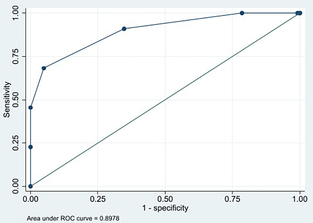
In this study, the HEALTH scoring system was calibrated using the Hosmer-Lemeshow goodness-of-fit test, which yielded a p-value of 0.896. This result indicates that the model’s estimates fit the data at an acceptable level and are well-calibrated. The discrimination ability of the HEALTH scoring system, based on the ROC curve, yielded a cut-off score of ≥4, with an AUC of 0.8978 (95% CI 0.823–0.971). The system demonstrated a sensitivity of 68.18% (95% CI 45.13%–86.14%) and a specificity of 95.04% (95% CI 89.52%–98.16%) ( Table 6 and Figure 1). Furthermore, the HEALTH scoring system was internally validated using the bootstrap resampling approach for 1,000 iterations. The Hosmer-Lemeshow test also produced a p-value of 0.896 in this validation, confirming that the scoring system has successfully passed internal validation.
The HEALTH scoring system was then applied to our data, dividing the subjects into two groups: those with a score of <4 (non-ITB) and those with a score of ≥4 (ITB), as detailed in Table 7. The HEALTH scoring system with a cut-off score of ≥4 successfully predicted 15 of 22 ITB cases, and a score of <4 successfully predicted 115 of 121 non-ITB cases. As a result, it yielded a positive predictive value of 71.43% (95% CI 52.14%–85.16%) and a negative predictive value of 94.26% (95% CI 89.90%–96.81%). The overall predictive accuracy and misprediction rate of this scoring system were 90.91% and 9.09%, respectively. The proposed HEALTH scoring system for differentiating ITB and non-ITB is presented in Table 8.
The HEALTH scoring system serves as a rule-in tool for assessing patients who exhibit clinical manifestations of ITB, which helps differentiate ITB from other gastrointestinal disorders. As illustrated in Figure 2, the system can be utilized when patients meet at least 3 out of 4 main clinical criteria and 1 of the 3 additional historical criteria. Subsequently, laboratory tests are performed using the HEALTH scoring system, which includes measurements of stool HBD-2 levels, ESR, blood ADA activity, lymphocytes, stool TB PCR, and NLR.
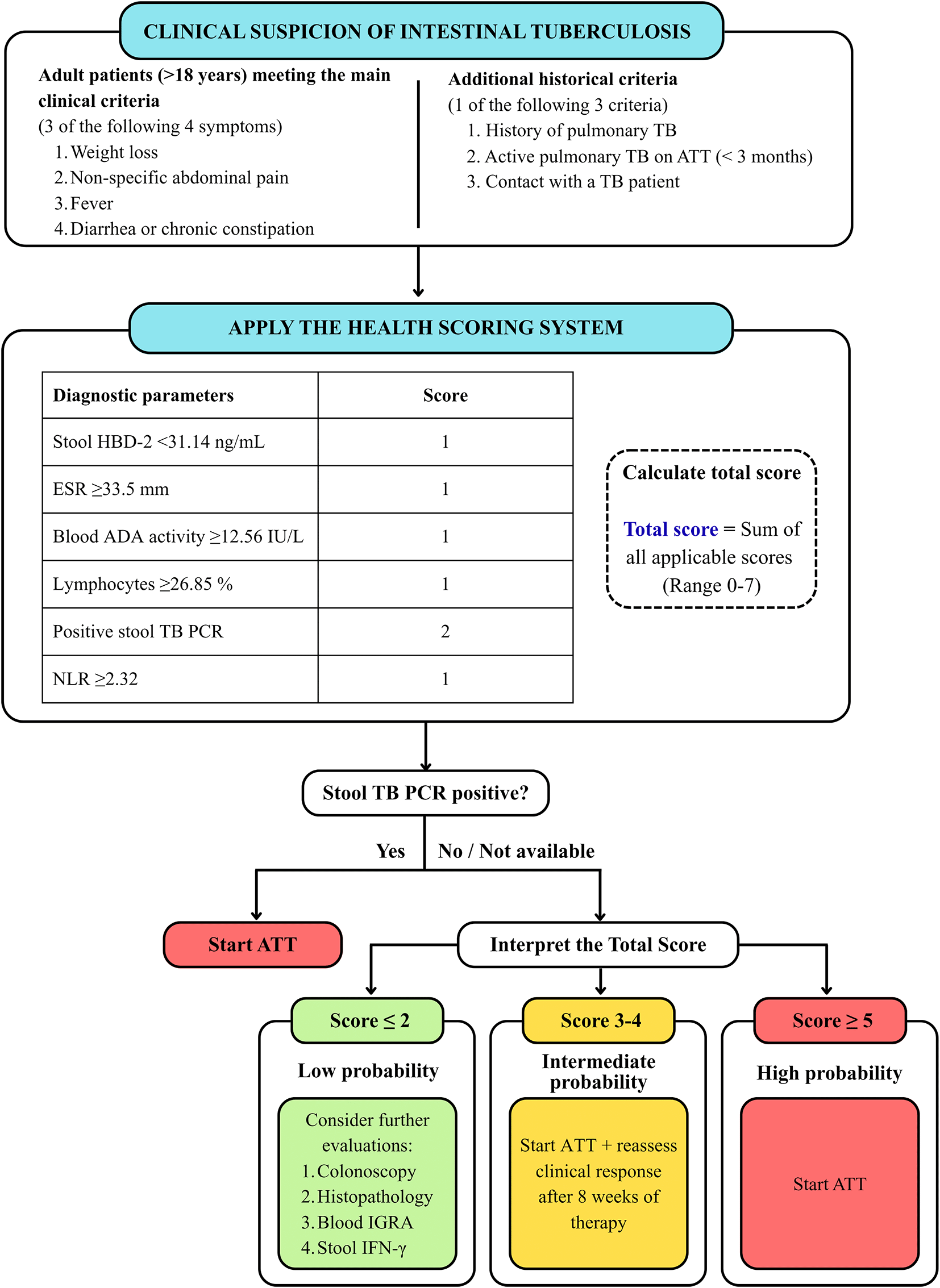
Patients with a positive stool TB PCR are considered to have microbiological evidence of ITB and are recommended to initiate ATT. For patients with a negative stool TB PCR result or when testing is unavailable, the total HEALTH score is interpreted as follows: (1) a score of ≤2 indicates a low probability of ITB and requires further diagnostic evaluation; (2) scores of 3–4 indicate an intermediate probability and support initiation of ATT, with a reassessment of clinical response after 8 weeks; and (3) a score of ≥5 indicates a high probability of ITB and supports the initiation of ATT.
Scoring systems in laboratory diagnostics play a crucial role in improving the accuracy and efficiency of disease diagnosis. By integrating specific biomarkers and clinical data, these systems enhance the ability to distinguish between similar diseases, predict disease progression, and guide treatment decisions. The development and validation of these systems across various diseases highlight their potential to transform clinical practice and improve patient outcomes. In Indonesia, a laboratory panel using a scoring system to diagnose ITB has not been developed.
There is currently no stand-alone laboratory test in ITB management, which creates opportunities for developing laboratory examinations using various biomarkers to diagnose ITB according to its pathophysiology. These examinations include routine examinations (such as hematology, IGRA, and blood ADA activity) and specific examinations (such as TB PCR). This study used Poisson regression and ROC curves to determine independent diagnostic parameters. After multivariate analysis, we developed and validated a combination of 6 biomarkers from blood and stool specimens to differentiate ITB and other gastrointestinal diseases, including stool HBD-2 level, ESR, blood ADA activity, lymphocytes, stool TB PCR, and NLR.
Stool HBD-2 levels in the ITB subjects were lower than non-ITB subjects. Researchers have noted limited studies on stool HBD-2 levels in ITB subjects. Several studies reported elevated stool HBD-2 levels in patients with other gastrointestinal diseases, such as inflammatory bowel disease and irritable bowel syndrome.26,27 Expression of HBD-2 can be stimulated through two pathways: stimulation by pro-inflammatory cytokines (e.g., IL-1β, IL-1α, and TNF-α) and direct recognition of microbial components (e.g., lipopolysaccharides, peptidoglycan, and flagellin) through specific receptors (e.g., IL-1R, TNF-R, and TLRs) and signaling pathways (NF-κB/AP-1/MAPK).18 The elevated stool HBD-2 levels observed in non-ITB subjects may therefore reflect persistent activation through both pathways. In addition, MTB possesses several immune response evasion strategies mediated by cell wall glycolipids, including phthicerol dimycocerosates (PDIM), which mask pathogen-associated molecular patterns, and sulfoglycolipids, which inhibit TLR-2 signaling.28,29 These mechanisms may reduce the effectiveness of host antimicrobial defenses and contribute to the distinct HBD-2 profile observed in ITB subjects, although the exact mechanisms regulating stool HBD-2 expression remain unclear.
The ESR in the ITB subjects was significantly higher compared to non-ITB subjects. These findings align with Zeng et al.30 and Hammami et al.,31 who reported that patients with ITB had higher ESR levels than their controls. The ESR test measures acute-phase proteins, which are inflammatory markers and can increase in a variety of diseases.30 In ITB, pro-inflammatory cytokines, particularly interleukin (IL)-6, IL-1, tumor necrosis factor-alpha (TNF-α), and IFN-γ, stimulate the liver to produce acute-phase proteins, including fibrinogen, resulting in the formation of erythrocyte rouleaux, which can elevate ESR results.32,33
Blood ADA activity in ITB subjects was significantly higher than non-ITB subjects. Studies evaluating blood ADA activity specific to patients with ITB remain scarce. Nevertheless, previous studies demonstrated elevated blood ADA activity in patients with EPTB.34,35 It is important to highlight that ADA activity measured in the blood does not pinpoint the exact production source but reflects the overall ADA activity from activated lymphocytes and monocytes throughout the body, including areas affected by MTB. Currently, ADA tests have not yet been validated on stool specimens or biopsy tissue, making it difficult to determine local ADA production in the intestine in ITB. The increased number of monocytes in ITB subjects correlates with the elevated ADA activity, suggesting that monocytes play a more significant role than other cells in the context of ITB.36,37
The percentage of lymphocytes in both ITB and non-ITB subjects showed no significant difference, remaining within the normal range (20–40%). These findings are comparable with those of Zeng et al., who reported a normal percentage of lymphocytes in patients with ITB.30 In TB infection, the percentage of lymphocytes in the blood often appears within the normal range due to complex mechanisms of immune responses. While some subsets of lymphocytes may change, the overall percentage of lymphocytes can remain within normal ranges. Infection with MTB can cause shifts in lymphocyte homeostasis; for example, subsets like CD8+ T-cells and B-cells may increase, while others like CD4+ T-cells may decrease. These changes can balance out, resulting in a normal overall percentage of lymphocytes.38,39
The proportion of positive stool TB PCR was significantly higher in the ITB subjects than non-ITB subjects. This study is similar to those reported by Suparmin et al.4 and may be influenced by the high endemicity of TB in the population. This study demonstrated “acceptable” specificity (100%) for the stool TB PCR test in differentiating ITB and non-ITB subjects. This finding aligns with Gaur et al., who reported that stool TB PCR tests showed high specificity in EPTB subjects.40 PCR is a recent advanced diagnostic method increasingly used to diagnose TB. Unlike biopsy, stool TB PCR is non-invasive and less prone to sampling error. The detailed mechanism of MTB DNA shedding in stool remains to be investigated. A recent study indicates a two-way interaction between the gut and lung (gut-lung axis) in the transfer of metabolites, immune cells, and bacterial fragments via the bloodstream.41 However, these findings require further investigation, as the mechanistic basis of ITB is still unclear. A major challenge in diagnosing ITB is its non-specific and diverse clinical manifestations, which delay confirmation. Therefore, detecting MTB DNA in ITB subjects’ stool may serve as a valuable rule-in test.
In the present study, the NLR, an indicator of acute infection or inflammation, remained within the normal range and showed no significant difference between ITB and non-ITB subjects. This finding contrasts with the results reported by Rees et al., who noted higher NLR levels in symptomatic pulmonary TB cases compared to controls.14 This discrepancy may be attributed to the role of neutrophils in non-granulomatous and acute inflammatory conditions. NRL is a newly identified biomarker of inflammation that is simpler to measure and relatively stable compared to other inflammatory biomarkers. However, the relationship between NLR and clinical outcomes can be influenced by several factors, including comorbidities, immune status, and virulence of the tuberculosis strain. Additionally, NLR can vary significantly between individuals based on age, gender, and ethnicity. It can also fluctuate over time due to stress, medications, and physiological conditions.
The HEALTH scoring system was developed and validated to help identify ITB in patients presenting with compatible gastrointestinal symptoms and a clinical history related to TB. Based on ROC analysis, the optimal cut-off was set at a score ≥4. This score exhibited a sensitivity of 68.18%, a specificity of 95.04%, and a strong LR+ of 13.75%. These results indicate that patients with a score of 4 or higher have a significantly increased probability of true ITB, and that LR+ values above 10 provide strong evidence that the HEALTH scoring system has potential as a rule-in tool for ITB in most circumstances.42 As a result, patients with these scores were managed according to the institutional ATT protocol.
However, despite the high specificity at the cut-off score of ≥4, depending only on this cut-off could result in missing several true ITB cases, as was the case with 7 of the 22 ITB subjects in this study, who may have had early symptoms, were paucibacillary (low bacterial counts), or were atypical. Therefore, patients with scores between 3 and 4 were classified as having an intermediate probability of having ITB, rather than being excluded outright. This decision is supported by the diagnostic performance observed at a lower cut-off (≥3), which showed a significantly higher sensitivity of 90.91% but a moderate specificity of 65.29%. This suggests that patients in the lower score range may still represent clinically significant cases of ITB. Consequently, individuals within the intermediate score category underwent additional clinical reassessment, follow-up evaluations after starting ATT, and further diagnostic investigations as needed. Meanwhile, scores of ≤2 were classified as low probability for ITB, initiating consideration of alternative diagnoses and further supportive testing, such as colonoscopy, histopathology, blood IGRA, and stool IFN-γ. This stratified approach aimed to balance diagnostic sensitivity and specificity while effectively integrating the likelihood ratio findings into practical clinical decision-making.
Developing laboratory panels using a scoring system based on subjects’ clinical biomarkers is nothing new. To manage the risk of patient severity or differentiate infectious diseases from one another, several previous studies have developed laboratory scoring systems, such as a lab score system for predicting COVID-19 patient severity, differentiating subjects with severe and mild H1N1 infection, and discriminating TB infection from both PTB and EPTB.43–45 However, researchers have pointed out that there are still very few reports regarding laboratory parameters for differentiating ITB from other gastrointestinal diseases. Makharia et al. developed a scoring system with 4 parameters based on clinical, endoscopic, and histological parameters to differentiate ITB and CD.20 Scoring system developed by Huang et al. combined 12 features based on laboratory, endoscopic, histologic, and radiographic features and showed that scores > −0.5 were diagnosed with CD; otherwise, patients were diagnosed with ITB.46 Wu et al. developed a predictive model for differentiating ITB from CD using 5 markers, including perianal disease, pulmonary involvement, longitudinal ulcer, left colon, and ratio of tuberculosis-specific antigen to phytohaemagglutinin.47 In addition, Qiu et al. developed a scoring system for differentiating colorectal ulcerative diseases, especially ITB, CD, and primary intestinal lymphoma, based on endoscopic ultrasound data.48
The strength of the HEALTH scoring system is that this is the first study based on laboratory findings that predicted the probability of ITB. Additionally, the laboratory tests involved in this scoring system are user-friendly and non-invasive, as they only require blood and stool samples. In Indonesia, this scoring system can be utilized by healthcare facilities, regardless of whether they have colonoscopy and histopathology services. Healthcare facilities that lack these services can use the HEALTH scoring system to assist clinicians in managing ITB. On the other hand, healthcare facilities equipped with colonoscopy and histopathology services can use the HEALTH scoring system to identify patients who should undergo colonoscopy, thereby reducing waiting times for colonoscopy examinations.
It should be noted that our study has several limitations. This study was a single-center cross-sectional study, the reagents used to examine stool IFN-γ in Indonesia were not included in the in vitro diagnostic reagents, and the model was only internally validated. Independent external validation, cost-effectiveness analysis, and a multicenter randomized prospective study must be designed to validate the HEALTH scoring system.
This study developed and validated a laboratory panel called the HEALTH scoring system based on clinical biomarkers of stool HBD-2 level, ESR, blood ADA activity, lymphocytes, stool TB PCR, and NLR, which could be used as a rule-in tool for differentiating ITB from other gastrointestinal diseases.
This study was approved by the Ethics Committee, Faculty of Medicine, Universitas Indonesia–Dr. Cipto Mangunkusumo National Central General Hospital (KET-1498/UN2.F1/ETIK/PPM.00.02/2020, Date: 10.12.2020 and ND-3/UN2.F1/ETIK/PPM.00.02/2022, Date: 10.01.2022). All study participants or participants’ family have given their written informed consent regarding their participation and data publication in this study.
The statistical analyses performed in this article using SPSS version 20 and STATA 14 software can be conducted using the freely accessible software R (programming language) https://www.r-project.org/.