Просмотр новости
Найдите то, что Вас интересует
 Спутники
 РН
РН SpaceX
 Роскосмос
 NASA
NASA
СпутникиРНSpaceXРоскосмосNASAIntroduction Invasive aspergillosis is a severe infection that usually affects immunocompromised patients. Primary cutaneous involvement is a rare presentation and presents a challenge for early diagnosis. We report a case of primary cutaneous aspergillosis (PCA) in an immunocompromised patient with no evident prior skin trauma and with pulmonary dissemination. Case report We report the case of primary cutaneous aspergillosis involving the left ankle in a 5-year-old girl with no history of preceding trauma. The patient was undergoing chemotherapy for lymphoblastic leukemia. Aspergillus hyphae were identified on skin biopsy. Cultures grew Aspergillus flavus. The diagnosis of cutaneous aspergillosis enabled the diagnosis of probable pulmonary aspergillosis, although there was no mycopathological proof of lung infection. The patient was treated with initial Amphotericin B followed by Voriconazole with complete skin and respiratory response. Conclusion This case underscores the critical need to consider cutaneous aspergillosis in immunocompromised patients with necrotic skin lesions— even in the absence of obvious trauma—as prompt diagnosis and treatment are vital to prevent dissemination and and improve outcomes.

KALBOUSSI Y, CHENBAH W, CHOUAIEB H et al. Case Report: Primary Cutaneous Aspergillosis in a Neutropenic Child with Pulmonary Dissemination: A case report [version 2; peer review: 2 approved, 1 approved with reservations]. F1000Research 2026, 15:454 (https://doi.org/10.12688/f1000research.178649.2)
Case Report
Revised
[version 2; peer review: 2 approved, 1 approved with reservations]
https://orcid.org/0009-0004-3616-2164
1,2, Wafa CHENBAH3,4, Hamed CHOUAIEB1,4, [...] Nour GAALOUL3,4, Samar ISMAILhttps://orcid.org/0000-0002-6152-0988
1,4, Monia GUERMAZI3,4, Mariem BEN TICHA4,5, Imene KHAMMARI1,4, Yosra BEN YOUSSEF3,4, Akila FATHALLAHhttps://orcid.org/0000-0002-4613-6689
1,4https://orcid.org/0009-0004-3616-2164
1,2, Wafa CHENBAH3,4, [...] Hamed CHOUAIEB1,4, Nour GAALOUL3,4, Samar ISMAILhttps://orcid.org/0000-0002-6152-0988
1,4, Monia GUERMAZI3,4, Mariem BEN TICHA4,5, Imene KHAMMARI1,4, Yosra BEN YOUSSEF3,4, Akila FATHALLAHhttps://orcid.org/0000-0002-4613-6689
1,41 Laboratory of Parasitology - Mycology, Farhat Hached University Hospital of Sousse, Sousse, Sousse, Tunisia
2 University of Monastir Faculty of Medicine of Monastir, Monastir, Monastir, Tunisia
3 Department of Hematology, Farhat Hached University Hospital of Sousse, Sousse, Sousse, Tunisia
4 University of Sousse Faculty of Medicine of Sousse, Sousse, Sousse, Tunisia
5 Department of Infectious diseases, Farhat Hached University Hospital of Sousse, Sousse, Sousse, Tunisia
Yasmine KALBOUSSI
Roles: Conceptualization, Data Curation, Investigation, Methodology, Writing – Original Draft Preparation, Writing – Review & Editing
Wafa CHENBAH
Roles: Conceptualization, Data Curation, Investigation, Methodology, Writing – Original Draft Preparation, Writing – Review & Editing
Hamed CHOUAIEB
Roles: Conceptualization, Methodology, Writing – Original Draft Preparation, Writing – Review & Editing
Nour GAALOUL
Roles: Conceptualization, Methodology, Writing – Original Draft Preparation, Writing – Review & Editing
Samar ISMAIL
Roles: Conceptualization, Methodology, Writing – Original Draft Preparation, Writing – Review & Editing
Monia GUERMAZI
Roles: Conceptualization, Methodology, Writing – Original Draft Preparation, Writing – Review & Editing
Mariem BEN TICHA
Roles: Conceptualization, Methodology, Writing – Original Draft Preparation, Writing – Review & Editing
Imene KHAMMARI
Roles: Conceptualization, Methodology, Writing – Original Draft Preparation, Writing – Review & Editing
Yosra BEN YOUSSEF
Roles: Validation, Visualization, Writing – Original Draft Preparation, Writing – Review & Editing
Akila FATHALLAH
Roles: Validation, Visualization, Writing – Original Draft Preparation, Writing – Review & Editing
OPEN PEER REVIEW
REVIEWER STATUS
Invasive aspergillosis is a severe infection that usually affects immunocompromised patients. Primary cutaneous involvement is a rare presentation and presents a challenge for early diagnosis. We report a case of primary cutaneous aspergillosis (PCA) in an immunocompromised patient with no evident prior skin trauma and with pulmonary dissemination.
Case reportWe report the case of primary cutaneous aspergillosis involving the left ankle in a 5-year-old girl with no history of preceding trauma. The patient was undergoing chemotherapy for lymphoblastic leukemia. Aspergillus hyphae were identified on skin biopsy. Cultures grew Aspergillus flavus. The diagnosis of cutaneous aspergillosis enabled the diagnosis of probable pulmonary aspergillosis, although there was no mycopathological proof of lung infection. The patient was treated with initial Amphotericin B followed by Voriconazole with complete skin and respiratory response.
ConclusionThis case underscores the critical need to consider cutaneous aspergillosis in immunocompromised patients with necrotic skin lesions— even in the absence of obvious trauma—as prompt diagnosis and treatment are vital to prevent dissemination and and improve outcomes.
Aspergillosis, cutaneous aspergillosis, neutropenia, invasive pulmonary aspergillosis, case report
Corresponding author: Yasmine KALBOUSSI Competing interests: No competing interests were disclosed.
Grant information: The author(s) declared that no grants were involved in supporting this work.
Copyright: © 2026 KALBOUSSI Y et al. This is an open access article distributed under the terms of the Creative Commons Attribution License, which permits unrestricted use, distribution, and reproduction in any medium, provided the original work is properly cited. How to cite: KALBOUSSI Y, CHENBAH W, CHOUAIEB H et al. Case Report: Primary Cutaneous Aspergillosis in a Neutropenic Child with Pulmonary Dissemination: A case report [version 2; peer review: 2 approved, 1 approved with reservations]. F1000Research 2026, 15:454 (https://doi.org/10.12688/f1000research.178649.2) First published: 30 Mar 2026, 15:454 (https://doi.org/10.12688/f1000research.178649.1) Latest published: 02 Jun 2026, 15:454 (https://doi.org/10.12688/f1000research.178649.2)
This revised version of the article has been substantially updated in response to peer review comments. Several clarifications have been made in the case presentation, including a more precise description of the timing of clinical events, particularly the interval between neutropenia, the appearance of cutaneous lesions, and the subsequent onset of pulmonary symptoms. Additional details have also been added regarding the initial diagnostic workup, including the absence of baseline imaging at presentation and the lack of respiratory sampling due to the patient’s clinical condition.
In the microbiological assessment, the interpretation of serum galactomannan has been revised to reflect its borderline positivity, and its role in the diagnostic process has been appropriately tempered.
The Discussion section has been significantly streamlined to remove redundant descriptions of the case and now focuses more directly on the main scientific message. Furthermore, the epidemiological context of invasive and cutaneous Aspergillus infections has been expanded.
Overall, these revisions improve the clarity, structure, and scientific rigor of the manuscript, while providing a more balanced interpretation of the diagnostic evidence and a clearer positioning of this case within the existing literature.
See the authors' detailed response to the review by Rim Ben Abdallah
See the authors' detailed response to the review by Rim Rakkez
Invasive aspergillosis is a severe and potentially fatal infection usually affecting immunocompromised patients.1,2 Pulmonary involvement is the predominant presentation, whereas cutaneous localization is much less frequent. This can develop as a primary infection, usually arising from direct inoculation of the skin3 or occur as part of a disseminated infection from the lung.1 The true incidence of primary cutaneous aspergillosis (PCA) among immunocompromised patients is not well established but seems to be rising, possibly as a result of better recognition and the increasing number of immunocompromised individuals.4,5 The major limitation in the management of these infections is the challenge of early diagnosis. We report a rare case of primary cutaneous aspergillosis (PCA) caused by Aspergillus flavus in a neutropenic and immunocompromised 5 year- old patient with no evident prior skin trauma. PCA is typically associated with breaches in skin integrity caused by catheters, dressings, or trauma, making the present case particularly unusual.
A 5-year-old girl was admitted to the Haematology department of Farhat-Hached hospital (Sousse, Tunisia) in September 2024 for acute lymphoblastic leukemia. The patient was treated according to EORTC protocole (Very High Risk aka VHR group) and received Chemotherapy VANDA consisting of : anthracyclines, L-asparginase, cyclophosphamide, methotrexate and corticosteroids (12mg of dexamethasone per day during 5 days). She had a cytological remission: 3% Blastic cells but a positive minimal residual disease (MRD) on the Last Bone Marrow assessement. In April 2025, she was admitted to receive consolidation chemotherapy. Seven days after the last dose, she developed high-grade fever (40 °C), concomitantly with the appearance of a small 5-mm black spot on her left ankle ( Figure 1), with no evidence of intravenous catheterization, local trauma, dressing, or other identifiable predisposing skin condition at the lesion site. The patient was neutropenic, with an absolute neutrophil count <500/mm3 for 5 days. Biological investigations demonstrated elevated inflammatory markers, including C-reactive protein (CRP), and bacterial and viral microbiological workup was negative. She was started on broad-spectrum antibiotics made of Piperacillin-Tazobactam and Vancomycin. Three days later, the patient still febrile and the skin lesion grew larger about 1.5 cm, becoming swollen with a crusted center and red surrounding skin. Antibiotics were switched to Imipenem and Ciprofloxacin with no improvement.
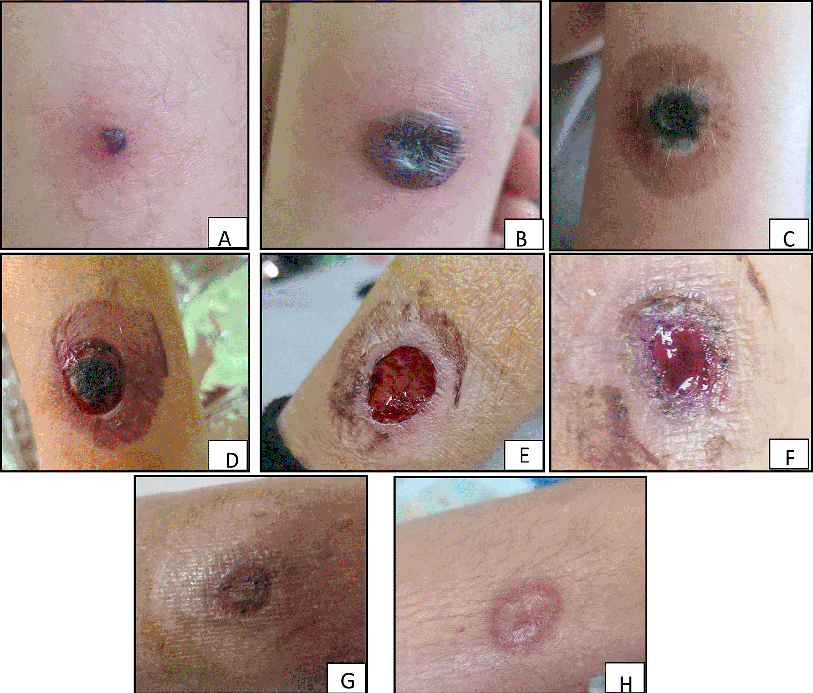
A, Initial skin lesion; B, Day 4; C, Day 5; D, Two days after Amphotericin; E, Day 7 of Amphotericin B; F, Lesion aspect after Voriconazole switch; G and H, Complete skin remission under Voriconazole.
When a mild cough appeared seven after the onset of the cutaneous lesion, a full-body CT scan was done. It showed nodules and lung infiltrates with a halo sign highly suggestive of angio-invasive pulmonary aspergillosis. Respiratory sampling was not performed due to the patient’s thrombocytopenia and clinical fragility and Intravenous amphotericin was started immediately. Within 12 hours, she became afebrile for the first time. The skin lesion on her ankle evolved: it became itchy, developed a necrotic center with a purplish halo, and later the center dried out, with a hemorrhagic edge and ulceration. Eventually, the necrotic center detached from the lesion. This necrotic tissue was sent to the mycology laboratory for analysis.
Microscopic examination revealed large, septate, and irregular hyphae with acute-angle branching, suggestive of Aspergillus ( Figures 2). The specimen was cultured on Sabouraud dextrose agar supplemented with chloramphenicol (SC) and incubated at 30 °C. Fungal growth, 4 days after incubation, was consistent with Aspergillus. Identification using the Vitek MS PRIME (bioMérieux, France), yielded Aspergillus flavus. Antifungal susceptibility testing was carried out using the MIC Test Strip method (Liofilchem, Roseto degli Abruzzi, Italy). The minimum inhibitory concentrations (MICs) were as follows: 0.75 mg/L for Amphotericin B and 0,38 mg/L for Voriconazole. No respiratory specimens were submitted for mycological examination. Concurrently, the serum galactomannan index was measured using the Platelia Aspergillus enzyme immunoassay (Bio-Rad, France), yielding a positive result with an index value of 0.57, using the manufacturer-recommended positivity cut-off of 0.5.
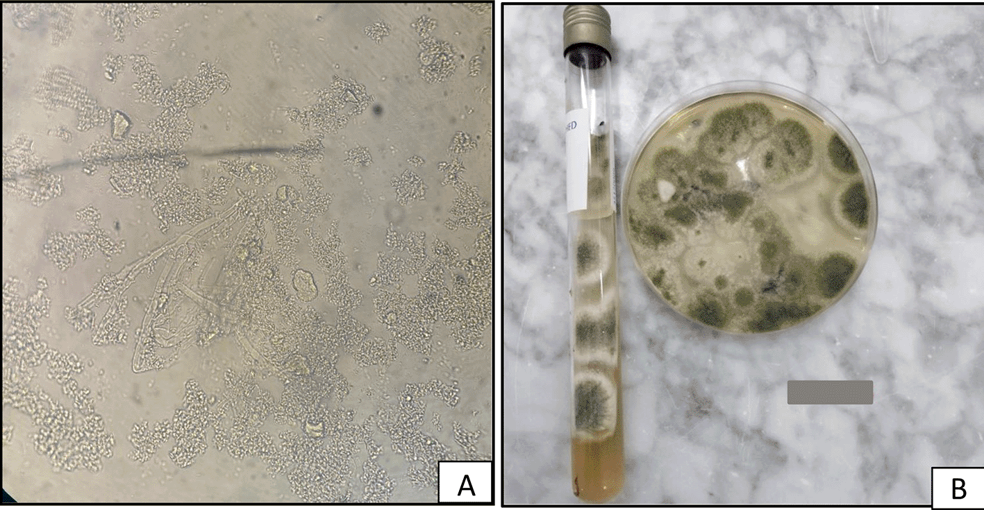
A, large, septate, and irregular hyphae with acute-angle branching on Direct Examination suggestive of Aspergillus (X100); B, Aspergillus flavus growing on culture.
Diagnosis of PCA with probable pulmonary dissemination was confirmed. Based on these findings, antifungal therapy was switched to Voriconazole, leading to complete resolution of the cutaneous lesions. The patient was subsequently discharged with a favorable outcome. With regular follow-up and local wound care, she achieved full recovery, and no relapse was observed at 6-month follow-up.
We report a rare case of primary cutaneous aspergillosis (PCA) due to Aspergillus flavus in a neutropenic patient with acute lymphoblastic leukemia, which subsequently progressed to invasive pulmonary aspergillosis. This case demonstrates the rapid progression of an initially inconspicuous cutaneous lesion to a severe necrotizing infection. It underscores the importance of careful clinical evaluation, as seemingly minor skin findings may represent an entry point for invasive fungal disease. Clinicians should maintain a high index of suspicion for primary cutaneous aspergillosis in neutropenic patients. Therefore, even a localized skin lesion requires aggressive systemic antifungal therapy to prevent dissemination.
Pediatric patients undergoing chemotherapy for hematological malignancies as illustrated by this 5-year-old girl, represent a high-risk cohort for invasive fungal infections.1,2 The population-based incidence of invasive aspergillosis (IA) in children remains unknown and is likely to vary between healthcare settings and countries. However, data from the Kids’ Inpatient Database, a large representative dataset of hospitalizations in the United States, reported an annual incidence of 437 cases per 100,000 (0.4%) hospitalized immunocompromised children in 2000 (new reference). The most associated disorders in children are leukemias and lymphomas.3 However, some cases have been reported with immunocompetent patients.4 Intensive regimens, including anthracyclines and cyclophosphamide, induce profound and prolonged neutropenia, which is the major risk factor for invasive aspergillosis.5 Corticosteroids, a key component of leukemia protocols, further impair immune defenses by suppressing macrophage and neutrophil function, crippling the host’s ability to contain fungal invasion. Consequently, this immunocompromised state, defined by cytotoxic and steroid-induced deficits, creates a perfect environment for invasive fungal diseases.
Cutaneous aspergillosis can occur either as a primary infection or as a secondary manifestation.6,7 Primary cutaneous aspergillosis (PCA) typically results from the direct inoculation of spores into the skin via breaches in barrier integrity, such as at catheter insertion sites, trauma wounds, or beneath occlusive dressings. In contrast, secondary cutaneous involvement occurs almost exclusively through hematogenous dissemination7,8 from a deep-seated focus, most commonly the lung. This form is associated with the angioinvasive behavior of Aspergillus species. Determining whether the infection is primary or secondary to a primary site, such as the lungs, is crucial for guiding treatment.9 In our case, the initial isolated cutaneous lesion, which appeared concomitantly with the onset of fever, and the absence of radiological lung abnormalities at presentation, strongly supports the diagnosis of primary cutaneous aspergillosis with subsequent pulmonary dissemination. However, we acknowledge the limitation that a secondary cutaneous involvement in the context of systemic disease cannot be completely excluded. This cutaneous aspergillosis occurred in the absence of any clinically apparent skin injury, which, to our knowledge, appears to be exceptional. However, a minor or unnoticed breach in the skin barrier cannot be excluded. This case highlights that primary cutaneous aspergillosis should be considered even in the absence of evident skin trauma.
PCA can have different presentations: erythematous macules and papules with pain and itching, necrotizing skin lesions, hemorragic bullas, ulcerations with central necrosis or violaceous nodules.10 The variety of presentations and the non-typical form of lesions lead to an underdiagnosis of PCA and emphasize on the importance of mycological examination in order to start antifungal treatment and avoid the dissemination.
The definitive diagnosis of cutaneous aspergillosis relies on examination and culture of a deep tissue biopsy, as superficial samples are often inadequate11 direct examination typically reveals septate hyphae with acute-angle branching, suggestive of Aspergillus, though not pathognomonic, as similar hyaline molds like Fusarium must be excluded.8 While A. fumigatus predominates in invasive aspergillosis overall, accounting for approximately 53% of pediatric cases in the largest multicenter study,1 A. flavus is notably prevalent in primary cutaneous infections (PCA), accounting for a significant proportion of cases.6,12 Our case aligns with this epidemiological profile for PCA. Serological biomarkers, such as serum galactomannan (GM), provide valuable adjunctive evidence. In pediatric patients, GM assay offers good sensitivity and specificity, and a positive result in a high-risk clinical context strongly supports the diagnosis of invasive disease.8 In this case, the galactomannan antigenemia (index 0.57) was borderline and not sufficient on its own to confirm disseminated infection.
According to the EORTC/MSGERC criteria,13 our patient fulfilled the definition of proven invasive cutaneous aspergillosis, based on the demonstration of septate hyphae in direct examination from a deep skin biopsy and confirmatory culture yielding Aspergillus. However, imaging alone is insufficiently specific for diagnosing pulmonary aspergillosis, as current guidelines require microbiological evidence for a probable infection.
The antifungal susceptibility testingc onfirmed a fully susceptible profile, with a notably low Voriconazole MIC of 0.38 mg/L, strongly justifying its use as primary therapy according to EUCAST guidelines. The patient’s sequential antifungal regimen—initial Amphotericin B followed by Voriconazole—is a common clinical strategy for managing suspected invasive fungal infections.14,15 Most patients treated with one or the other had full recovery.16 However, this approach requires careful consideration due to on going debates about potential antagonistic interactions between the two drug classes.17 Surgical debridementand oral Itraconazole are also therapeutic options with extended lesions.18
This clinical case highlights the importance of considering cutaneous aspergillosis in immunocompromised patients presenting with skin lesions that progress to necrosis, even in the absence of trauma history. Particular attention must be paid to such lesions, as prompt diagnosis and treatment are crucial to prevent dissemination and reduce infection-related mortality.
A written consent was provided and signed by the patient’s parent, including the authorization for publishing clinical details and/or clinical images.
The CARE chechlist is publicly available at Zenodo.19
Title: Completed CARE checklist associated with the manuscript entitled “Primary Cutaneous Aspergillosis in a Neutropenic Child with Pulmonary Dissemination: A case report”.
DOI: https://doi.org/10.5281/zenodo.18860965.19
License: CC0 1.0.
The authors would like to thank the laboratory technicians and nursing staff for their valuable contribution to the patient’s care.
The author(s) declared that no grants were involved in supporting this work.
© 2026 KALBOUSSI Y et al. This is an open access article distributed under the terms of the Creative Commons Attribution License, which permits unrestricted use, distribution, and reproduction in any medium, provided the original work is properly cited.
Current Reviewer Status: ?
Key to Reviewer Statuses VIEW HIDE
ApprovedThe paper is scientifically sound in its current form and only minor, if any, improvements are suggested
Approved with reservations A number of small changes, sometimes more significant revisions are required to address specific details and improve the papers academic merit.
Not approvedFundamental flaws in the paper seriously undermine the findings and conclusions
Version 2
VERSION 2
PUBLISHED 02 Jun 2026
Revised
Reviewer Report 24 Jul 2026
Christopher Heath, Department of Medicine, University of Western Australia, Perth, Western Australia, Australia; Infectious Diseases & Clinical Microbiology, Fiona Stanley Hospital, Murdoch, Western Australia, Australia
Approved with Reservations
VIEWS 0
Is the background of the case’s history and progression described in sufficient detail?
Partly
Are enough details provided of any physical examination and diagnostic tests, treatment given and outcomes?
Partly
Is sufficient discussion included of the importance of the findings and their relevance to future understanding of disease processes, diagnosis or treatment?
Partly
Is the case presented with sufficient detail to be useful for other practitioners?
Yes
References
1. Dutkiewicz M, Garros M, Bui J, Charlier V, et al.: Comparison of MALDI-TOF MS instruments and databases for the identification of uncommon yeasts,Aspergillus spp. and rare filamentous fungi. Journal of Clinical Microbiology. 2025; 63 (6). Publisher Full TextCompeting Interests: No competing interests were disclosed.
Reviewer Expertise: Clinical and Laboratory Mycology, Infections in the Immunocompromised Host (e.g., BM transplant medicine), Infections after solid organ transplantation, major burns and major trauma, and Infections in the ICU.
CloseReviewer Report 21 Jul 2026
Rim Ben Abdallah, Institut Pasteur de Tunis, Place Pasteur, Tunisia
Approved
VIEWS 0
Competing Interests: No competing interests were disclosed.
Reviewer Expertise: parasitology-mycology
CloseReviewer Report 09 Jun 2026
Rim Rakkez, Hematology, University Hospital of Martinique (Ringgold ID: 55525), Fort-de-France Bay, Fort-de-France, Martinique
Approved
VIEWS 0
Competing Interests: No competing interests were disclosed.
Reviewer Expertise: acute leukemias, myeloid malignancies
CloseVersion 1
VERSION 1
PUBLISHED 30 Mar 2026
Reviewer Report 18 May 2026
Rim Rakkez, Hematology, University Hospital of Martinique (Ringgold ID: 55525), Fort-de-France Bay, Fort-de-France, Martinique
Approved
VIEWS 0
Is the background of the case’s history and progression described in sufficient detail?
Partly
Are enough details provided of any physical examination and diagnostic tests, treatment given and outcomes?
Yes
Is sufficient discussion included of the importance of the findings and their relevance to future understanding of disease processes, diagnosis or treatment?
Yes
Is the case presented with sufficient detail to be useful for other practitioners?
Yes
Competing Interests: No competing interests were disclosed.
Reviewer Expertise: acute leukemias, myeloid malignancies
CloseReviewer Report 11 Apr 2026
Rim Ben Abdallah, Institut Pasteur de Tunis, Place Pasteur, Tunisia
Approved with Reservations
VIEWS 0
Is the background of the case’s history and progression described in sufficient detail?
Yes
Are enough details provided of any physical examination and diagnostic tests, treatment given and outcomes?
Partly
Is sufficient discussion included of the importance of the findings and their relevance to future understanding of disease processes, diagnosis or treatment?
Partly
Is the case presented with sufficient detail to be useful for other practitioners?
Partly
Competing Interests: No competing interests were disclosed.
Reviewer Expertise: parasitology-mycology
CloseOpen Peer Review
Alongside their report, reviewers assign a status to the article:
| Invited Reviewers | |||
|---|---|---|---|
| 1 | 2 | 3 | |
| Version 2 (revision) 02 Jun 26 | read | read | read |
| Version 1 30 Mar 26 | read | read | |
Sign up for content alerts
Alongside their report, reviewers assign a status to the article:
Approved - the paper is scientifically sound in its current form and only minor, if any, improvements are suggested
Approved with reservations - A number of small changes, sometimes more significant revisions are required to address specific details and improve the papers academic merit.
Not approved - fundamental flaws in the paper seriously undermine the findings and conclusions