Просмотр новости
Найдите то, что Вас интересует
 Спутники
 РН
РН SpaceX
 Роскосмос
 NASA
NASA
СпутникиРНSpaceXРоскосмосNASAFrom fluorescent ferns to sprawling neurons, the 2025 Nikon Small World contest showcased the artistry of life as seen through a microscope.
Through the lens of a microscope, even a dirty windowsill can hold treasures.
The rice weevil (Sitophilus oryzae) is a pest that devours grains and seeds. Seen atop a grain of rice, this glamour shot, above, won first place in the 2025 Nikon Small World photomicrography competition. Zhang You stumbled upon the dead insect while cleaning his house. He captured the rice weevil’s final flight by stacking more than 100 photos taken with a microscope.
Finding a dead weevil with its wings outstretched was lucky. “Their tiny size makes manually preparing spread-wing specimens extremely difficult,” says You. “So this naturally preserved individual is rare.” You’s a member of the Entomological Society of China. (Entomologists study insects.)
He mounted this weevil on a grain of rice to highlight its small size. He hopes the photo can provide insights into the pest’s structure. “To me, a standout work blends artistry with scientific rigor,” he says of the image. It captures “the very essence, energy and spirit of these creatures.”
The perched rice weevil was one of 71 striking photos honored on October 15 in this year’s competition. Here are a few of our other favorites.
A drop bursting with life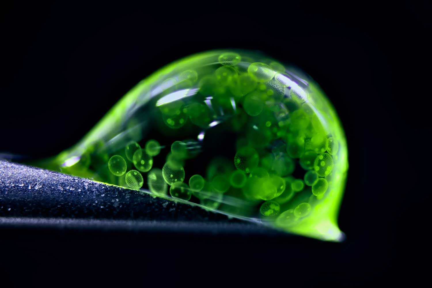 Jan Rosenboom
This orb — a water droplet — hosts a multi-celled universe.
Jan Rosenboom photographed hundreds of algae cells moving through the droplet. The German photographer and chemist used LED lights to capture the image. He also used a 50-year-old microscope, one that he purchased at age 14.
The algae here (Volvox) bloom in freshwater ponds, lakes and puddles. Their oval-shaped cells are outlined in green. The brighter green polka dots inside the outlines are young algae forming within each mother cell.
The droplet rests at a precarious angle on the tip of a syringe. Rosenboom spilled more than 20 droplets before snapping this second-place photo.
This image shows how much life can exist in a single drop of water, Rosenboom says. He hopes it helps people understand that even small worlds have biodiversity worth protecting.
Fluorescent ferns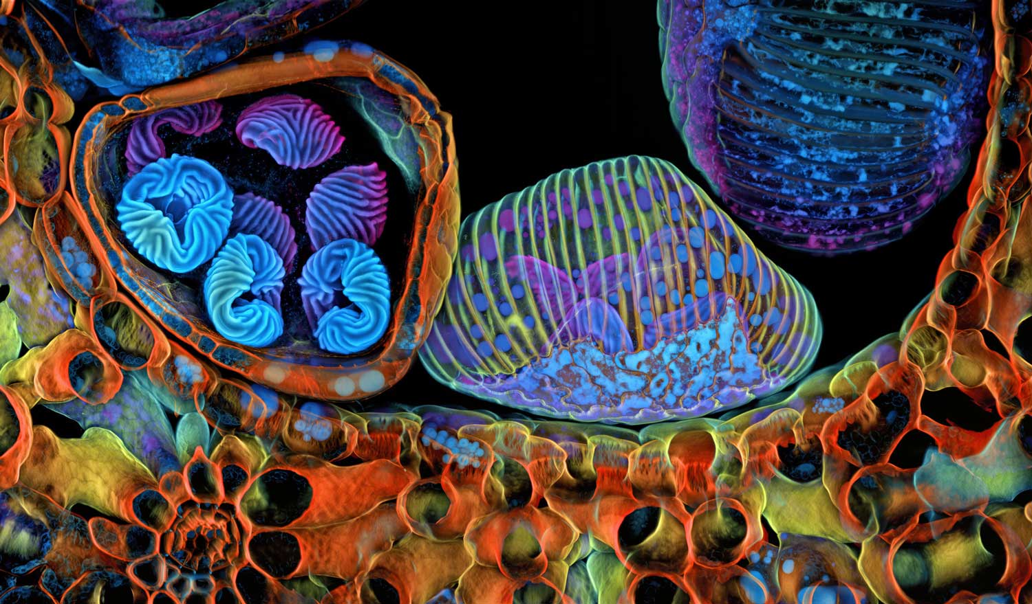 Igor Robert Siwanowicz
Kaleidoscopic pop art? No, these are the spores of a humble fern, shown in vibrant detail. Spores are cells that some plants, fungi and bacteria use to reproduce. Like seeds, they’re often spread by the wind or small animals.
Igor Siwanowicz lit these here using fluorescent lasers in a confocal microscope. This type of microscope shines lasers at a specimen to get rid of out-of-focus light. One advantage: It produces crisp images.
This image shows three podlike structures. Known as sporangia, these structures are where a fern’s spores grow. Siwanowicz sliced the pods in half with a small razor. This revealed what was inside them. The left pod holds spores with brain-like shells. The center pod remains unopened. And the right pod is empty. It likely burst and dropped its spores while he was prepping for the photo, Siwanowicz says. The frond tissue of the surrounding fern (Ceratopteris richardii) is orange.
Siwanowicz studies anatomy at the Howard Hughes Medical Institute’s Janelia Research Campus. That’s in Lansdowne, Va. He especially enjoys the chaos of the image, which won fifth place.
“Microscopy images are very abstract and sometimes alien-looking,” says Siwanowicz. “They’re confusing. But sometimes confusion creates this urge to find out more.”
Sprawling neurons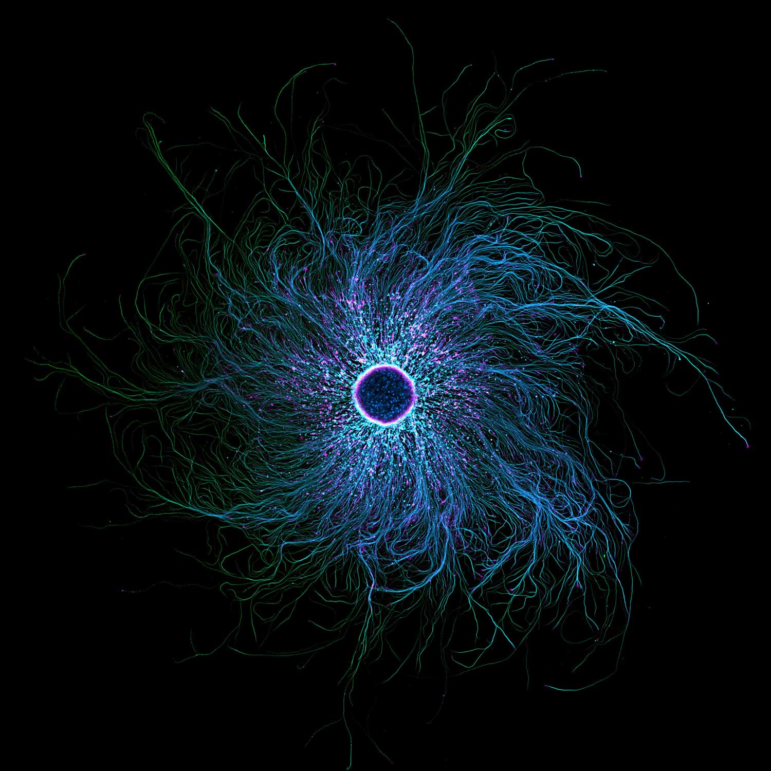 Stella Whittaker
This web of neurons may resemble the iris of an eye. But their spindly structures are designed to enhance touch, not sight.
Those wispy, rootlike arms help people sense what’s around them, says Stella Whittaker. A biophysicist, she studies the structure of sensory neurons. She took this photo while working at the National Institute of Neurological Disorders and Stroke in Bethesda, Md.
Her image displays the swirling branches of a type of neurons found in our arms, legs and spine. Whittaker colored the image with fluorescent molecule tags. These bind to proteins in the neurons. When the microscope’s laser hits the tag, it causes the neurons to light up.
The pink, speckled dots highlight actin. It’s a protein that helps muscles contract. The blue and green bits are microtubules. These act as highways for transporting organelles in and out of cells. At the center, 10,000 cells group together.
Studying the structure of neurons can help researchers understand how diseases develop in the brain, Whittaker explains. She’s now earning her Ph.D. at Weill Cornell School of Medicine in New York City.
Hairy forest?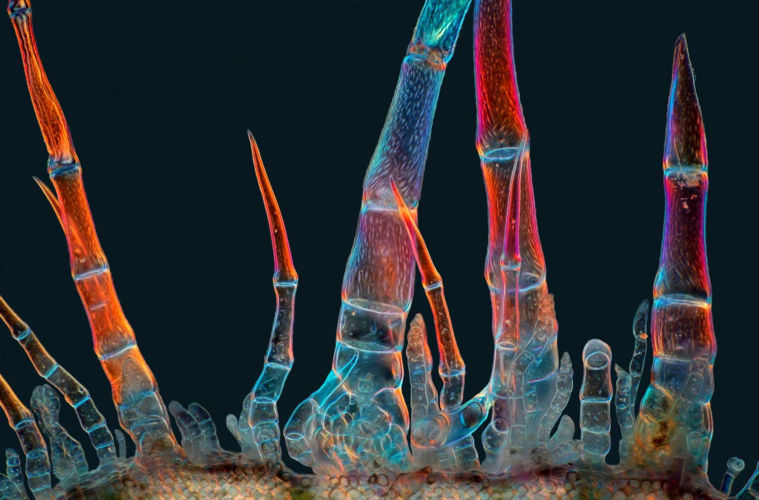 Marek Miś
This may look like a chorus line of insect legs reaching for the sky. In fact, these are trichomes (TRY-kohms). Such super-tiny structures protrude from the surface of many plants. They provide some sort of physical or toxic chemical defense against pests or disease-causing agents.
The ones shown here were found on sunflowers. Marek Miś couldn’t resist sticking slices of this bristly tissue under his microscope. Miś is a photographer and longtime nature-lover based in Poland.
Submit your question here, and we might answer it an upcoming issue of Science News Explores
He collected the trichomes from a bloom’s stem. He then lit them up with light passed through a homemade polarizing filter. Only light waves wiggling at certain angles could pass through the filter. That changed the hue these trichomes reflected.
The mesmerizing magentas and blues in the final image are stitched together from more than 100 photos. They highlight how trichomes come in a variety of shapes and sizes.
“Trichomes are not all the same,” Miś says. “The sunflower, already a beautiful plant in itself, hides additional beauty that cannot be seen with the naked eye.”
Tiny intestines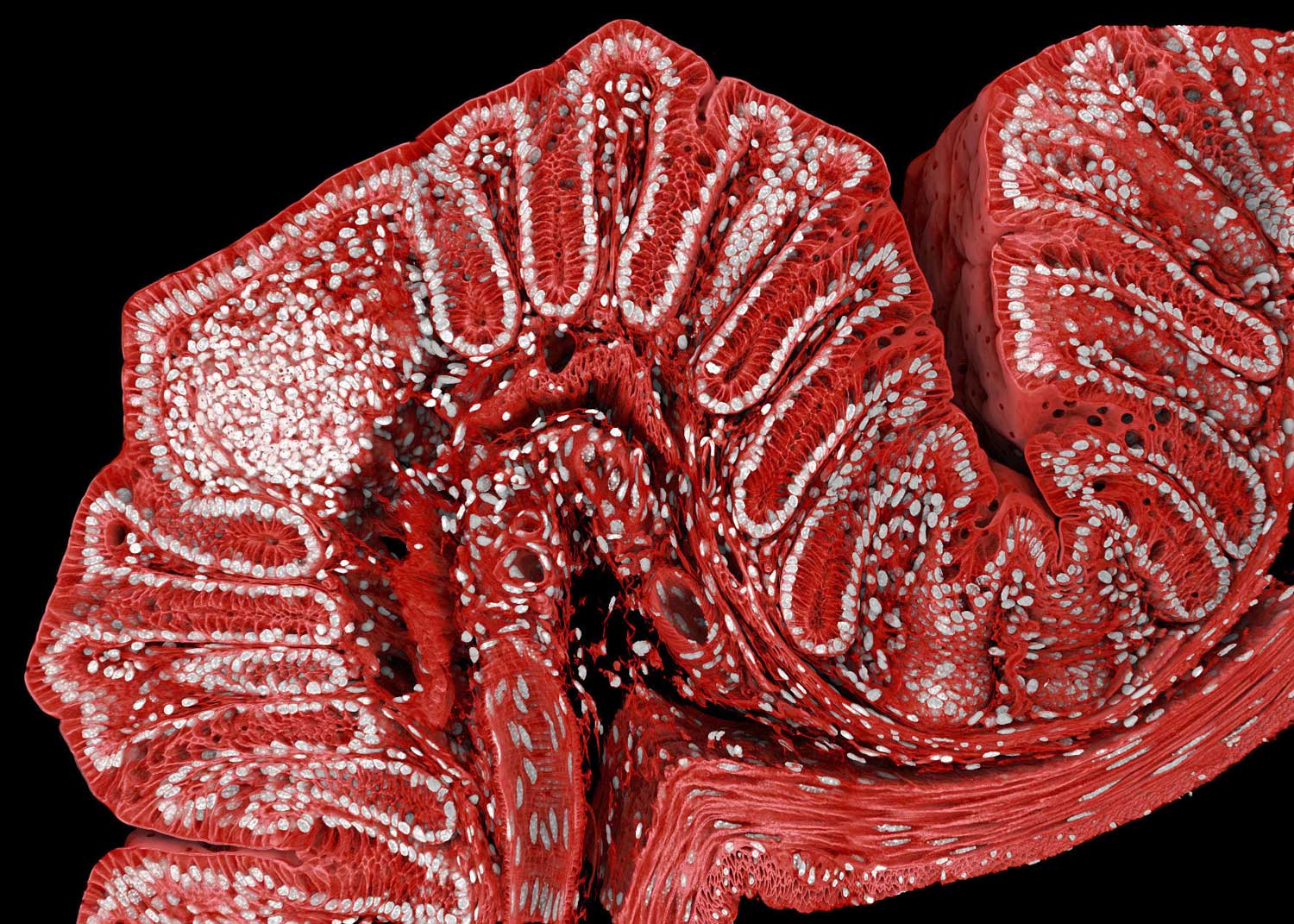 Marius Mählen, Koen Oost, Prisca Liberali, Laurent Gelman
What may resemble the decorative applique on some fashionable gown is actually the closeup of a mouse colon. It certainly brings glamour to the gut.
Marius Mählen is part of a team that wanted to learn how tissues grow. “If you figure out the rules by which healthy tissue forms,” he explains, ”then you will also start to understand why cancer tumors, for example, form very weird-shaped tissues.”
Mählen is a biologist at the Friedrich Miescher Institute for Biomedical Research. That’s in Basel, Switzerland. There, his team studies organelles in mouse cells. Their goal is to see how diseases, such as colon cancer, develop.
Over the course of one weekend, the team took 600 images of one mouse colon. They used a confocal microscope, then stitched the images together to create the final image.
By adding antibodies and dye, Mählen and his team marked the cells in the image with different colors. The nuclei of the cells appear in white. The cell membranes are now red. And the white clump toward the left marks a horde of immune cells scouting for invaders.
actin: A protein that can form long chains in cells. In muscle cells, these chains help the muscle contract. Actin chains also help cells move.
algae: A group of single-celled and multicellular organisms, once considered plants (they aren’t). As aquatic organisms, they grow in water. Like green plants, they depend on sunlight to make their food.
anatomy: (adj. anatomical) The study of the organs and tissues of animals. Or the characterization of the body or parts of the body on the basis of structure and tissues. Scientists who work in this field are known as anatomists.
angle: The space (usually measured in degrees) between two intersecting lines or surfaces at or close to the point where they meet.
antibodies: Any of a large number of proteins that the body produces from B cells and releases into the blood supply as part of its immune response. The production of antibodies is triggered when the body encounters an antigen, some foreign material. Antibodies then lock onto antigens as a first step in disabling the germs or other foreign substances that were the source of those antigens.
bacteria: (singular: bacterium) Single-celled organisms. These dwell nearly everywhere on Earth, from the bottom of the sea to inside other living organisms (such as plants and animals). Bacteria are one of the three domains of life on Earth.
biodiversity: (short for biological diversity) The number and variety of species found within a localized geographic region.
biomedical: Having to do with medicine and how it interacts with cells or tissues.
cancer: Any of more than 100 different diseases, each characterized by the rapid, uncontrolled growth of abnormal cells. The development and growth of cancers, also known as malignancies, can lead to tumors, pain and death.
cell: (in biology) The smallest structural and functional unit of an organism. Typically too small to see with the unaided eye, it consists of a watery fluid surrounded by a membrane or wall. Depending on their size, animals are made of anywhere from thousands to trillions of cells. Most organisms, such as yeasts, molds, bacteria and some algae, are composed of only one cell.
cell membrane: A structure that separates the inside of a cell from what is outside of it. Some particles are permitted to pass through the membrane.
chaos: A situation that is confused and has no order.
colon: (in biology) The majority of the large intestine, it runs between the cecum (a pouch below the small intestine) and the rectum. Foods are not digested in the colon, although this tissue lubricates wastes that will be excreted. Some liquids and salts, however, will be removed from materials stored in the colon before excretion.
contract: To activate muscle by allowing filaments in the muscle cells to connect. The muscle becomes more rigid as a result.
disorder: (in medicine) A condition where the body does not work appropriately, leading to what might be viewed as an illness. This term can sometimes be used interchangeably with disease.
filter: (n.) Something that allows some materials to pass through but not others, based on their size or some other feature. (v.) The process of screening some things out on the basis of traits such as size, wavelength, density, electric charge.
fluorescent: (v. fluoresce) Adjective for something that is capable of absorbing and reemitting light. That reemitted light is known as fluorescence.
freshwater: A noun or adjective that describes bodies of water with very low concentrations of salt. It’s the type of water used for drinking and making up most inland lakes, ponds, rivers and streams, as well as groundwater.
frond: The large leaf (or leaf-like) part of a fern, palm or other plant whose edges have many parts to it, sometimes known as leaflets.
fungi: (sing: fungus) Organisms with one or more cells that reproduce via spores and feed on living or decaying organic matter. Examples include mold, yeasts and mushrooms.
gut: An informal term for the gastrointestinal tract, especially the intestines.
immune: (adj.) Having to do with immunity. (v.) Able to ward off a particular infection. Alternatively, this term can be used to mean an organism shows no impacts from exposure to a particular poison or process. More generally, the term may signal that something cannot be hurt by a particular drug, disease or chemical.
infest: To create a parasitic community, such as when wasps infest the porch of an abandoned house. Such a community of pests is known as an infestation.
inflammatory bowel disease.: (IBD) A term for several potentially severe conditions that regularly (chronically) inflame the digestive tract. The two most common types of this disease are ulcerative colitis (which inflames the large intestine, or colon) and Crohn’s disease (which inflames the entire digestive tract). Both conditions stem from the immune system's abnormal response to the affected tissues. That means they are autoimmune diseases. Symptoms can include diarrhea, blood in the stool, abdominal pain and extreme fatigue.
insect: A type of arthropod that as an adult will have six segmented legs and three body parts: a head, thorax and abdomen. There are hundreds of thousands of insects, which include bees, beetles, flies and moths.
insight: The ability to gain an accurate and deep understanding of a situation just by thinking about it, instead of working out a solution through experimentation.
iris: The colored part of the eye, surrounding the pupil. The iris is a muscle.
laser: A device that generates an intense beam of coherent light of a single color. Lasers are used in drilling and cutting, alignment and guidance, in data storage and in surgery.
LED: (short for light emitting diode) Electronic components that, as their name suggests, emit light when electricity flows through them. LEDs are very energy-efficient and often can be very bright. They have lately been replacing conventional lights for home and commercial lamps.
membrane: A barrier which blocks the passage (or flow through) of some materials depending on their size or other features. Membranes are an integral part of filtration systems. Many serve that same function as the outer covering of cells or organs of a body.
microscope: An instrument used to view objects — such as bacteria or the single cells of plants or animals — that are too small to be visible to the unaided eye.
multicellular: Having or consisting of many cells. This includes all animals and plants, and many types of fungus.
muscle: A type of tissue used to produce movement by contracting its cells, known as muscle fibers. Muscle is rich in protein, which is why predatory species seek prey containing lots of this tissue.
neurological: An adjective that refers to the brain, spinal cord or nerves.
neuron: The main cell type of the nervous system — the brain, spinal column and nerves. These specialized cells transmit information by producing, receiving and conducting electrical signals. Neurons also can transmit signals to other cells with chemical messengers.
orb: Something having a spherical shape.
organelle: Specialized structures, such as mitochondria, found within a cell.
protein: A compound made from one or more long chains of amino acids. Proteins are an essential part of all living organisms. They form the basis of living cells, muscle and tissues; they also do the work inside of cells. Antibodies, hemoglobin and enzymes are all examples of proteins. Medicines frequently work by latching onto proteins.
sporangia: (sing. sporangium) Pod-like structures in which form a fern’s spores reproductive cells, known as spores.
spore: A tiny, typically single-celled body that is formed by certain bacteria in response to bad conditions. Or it can be the single-celled reproductive stage of a fungus (functioning much like a seed) that is released and spread by wind or water. Most are protected against drying out or heat and can remain viable for long periods, until conditions are right for their growth.
syringe: A plunger-type device used to extract blood from the body or to deliver a liquid medicine through the skin via a needle.
tissue: Made of cells, it is any of the distinct types of materials that make up animals, plants or fungi. Cells within a tissue work as a unit to perform a particular function in living organisms. Different organs of the human body, for instance, often are made from many different types of tissues.
trichome: A fine, hair-shaped structure that grows out from the surface of several types of plant parts, such as leaves, stems and fruits. They help defend their host plant from things that might stress it. Such stressors include the sun’s ultraviolet radiation, extreme temperatures, herbivores and disease-causing agents. Some trichomes, for instance, can store or make toxic chemicals to deter insects or other predators.
wave: A disturbance or variation that travels through space and matter in a regular, oscillating fashion.
weevil: Small beetles that belong to the Curculionidae family. There may be at least 60,000 different species. They tend to have a snout-like head part that allows them to penetrate seeds, nuts and plant stems.
A version of this article appears in the April 1, 2026 issue of Science News Explores.
About Carly Kay
Carly Kay is the Fall 2025 science writing intern at Science News. She holds a bachelor’s degree in communication from the University of California, Santa Barbara and a master’s degree in science communication from the University of California, Santa Cruz.