Просмотр новости
Найдите то, что Вас интересует
 Спутники
 РН
РН SpaceX
 Роскосмос
 NASA
NASA
СпутникиРНSpaceXРоскосмосNASAThis study describes tissue culture protocols for plantlet regeneration through indirect somatic embryogenesis and indirect organogenesis of Dorema microcarpum Korov. and Dorema sabulosum Litv. (Apiaceae) for the first time. The highest percentage of callusogenesis (90%) was obtained by using 2,4 -D (0.2 and 0.5 mg/l), followed by 2,4 -D (0.5 mg/l) + TDZ (0.5 mg/l) and 2,4-D (0.5 m g/l) + Kin (0.5 mg/l). Additionally, IAA (0.2 mg/l) + BAP (0.5 mg/l) showed the high percentage of callusogenesis (60–70%) for both Dorema species. In D. microcarpum, the highest percentage of callusogenesis was obtained from 2,4-D (0.5 mg/l) + TDZ (0.5 mg/l), while the highest percentage of embryogenesis was achieved with 2,4-D (0.5 mg/l) + Kin (0.5 mg/l). Hypocotyl explants yielded the highest percentage of embryogenesis. For somatic embryo maturation, 2,4-D had to be removed from a nutrient medium. Embryo maturation occurs after the embryo was transferred to a hormone-free medium. For D. sabulosum, morphogenesis occured through indirectly organogenesis, and the most optimal combination for the process of hemogenesis was IAA (0.5 mg/l) + BAP (1.0 mg/l). In vitro propagation of Dorema species in the future will become the basis for continuous year-round propagation of rare, endemic and medicinal species of this genus by biotechnological methods, and will become an alternative to obtaining biologically active compounds of medicinal plants.
Study Protocol
[version 1; peer review: awaiting peer review]
https://orcid.org/0000-0003-2035-7560
1, Mohizar Mirzaolimova2, Amir Zarekarizi3, [...] Feruza Mustafina4, Vasila Sharipova5, Khilola Ubaydullayeva6, Mukhayyo Kholdorova7, Gulsauir Kurbaniyazovahttps://orcid.org/0000-0003-0103-0298
8, Mirzohid Mirzaolimov9, Huseyin Turker10, Ziyoviddin Yusupov11, Komiljon Tojibaev12https://orcid.org/0000-0003-2035-7560
1, Mohizar Mirzaolimova2, [...] Amir Zarekarizi3, Feruza Mustafina4, Vasila Sharipova5, Khilola Ubaydullayeva6, Mukhayyo Kholdorova7, Gulsauir Kurbaniyazovahttps://orcid.org/0000-0003-0103-0298
8, Mirzohid Mirzaolimov9, Huseyin Turker10, Ziyoviddin Yusupov11, Komiljon Tojibaev121 Institute of Botany of Academy of Sciences of the Republic of Uzbekistan, Tashkent, 100125, Uzbekistan
2 Department of Biology, Faculty of Biotechnology, Namangan State University, Namangan, Uzbekistan
3 Department of Botany, University of Otago, Dunedin, New Zealand
4 The Botanical garden of the Republic of Uzbekistan, Tashkent, 100125, Uzbekistan
5 Institute of Botany of Academy of Sciences of the Republic of Uzbekistan, Tashkent, 100125, Uzbekistan
6 Department of Microbiology, Virology and Immunology, Bukhara State Medical Institute, Bukhara, Uzbekistan
7 6Jizzakh State pedagogical university, Jizzakh, Uzbekistan
8 Institute of Botany of Academy of Sciences of the Republic of Uzbekistan, Tashkent, 100125, Uzbekistan
9 Department of General Medical Sciences, Faculty of Medicine, Namangan State University, Namangan, Uzbekistan
10 Biotechnology Department, Faculty of Sciences, Niğde Ömer Halisdemir University, Nigde, Turkey
11 Institute of Botany of Academy of Sciences of the Republic of Uzbekistan, Tashkent, 100125, Uzbekistan
12 Institute of Botany of Academy of Sciences of the Republic of Uzbekistan, Tashkent, 100125, Uzbekistan
Dilafruz Jamalova
Roles: Conceptualization, Investigation, Methodology, Writing – Original Draft Preparation
Mohizar Mirzaolimova
Roles: Conceptualization, Data Curation, Investigation, Methodology, Resources
Amir Zarekarizi
Roles: Conceptualization, Formal Analysis, Software, Validation
Feruza Mustafina
Roles: Data Curation, Formal Analysis, Methodology, Supervision
Vasila Sharipova
Roles: Conceptualization, Formal Analysis, Validation
Khilola Ubaydullayeva
Roles: Conceptualization, Writing – Review & Editing
Mukhayyo Kholdorova
Roles: Data Curation, Formal Analysis, Investigation
Gulsauir Kurbaniyazova
Roles: Conceptualization, Investigation, Methodology
Mirzohid Mirzaolimov
Roles: Data Curation, Software, Validation
Huseyin Turker
Roles: Validation, Writing – Review & Editing
Ziyoviddin Yusupov
Roles: Project Administration, Supervision
Komiljon Tojibaev
Roles: Formal Analysis, Project Administration, Supervision
OPEN PEER REVIEW
REVIEWER STATUS AWAITING PEER REVIEW
The Apiaceae Lindl. family, one of the largest and most diverse plant families in the Angiosperm group, widely distributed across temperate regions, particularly in Central Asia, and highly valuable with its phytochemical content, essential oils and coumarins (Mottaghipisheh et al., 2021), some representatives are used and grown for household and medicinal purposes (Asnaashari et al., 2011; Dehghan et al., 2009; Amirkhiz et al., 2013). Within this family, the genus Dorema D. Don represents a small but ecologically and pharmacologically significant group, comprising approximately 12 accepted worldwide species (http://www.theplantlist.org). Dorema species are characterized as monocarpic perennials, blooming only once during life cycle and propagating by seeds (Pimenov, 1988; Nechaeva, 1985) This reproductive strategy, combined with their limited distribution, overharvesting and specific habitat requirements, put many Dorema species potentially endangered and vulnerable to environmental changes and anthropogenic pressures (Amiri, 2016). The genus has been used as a food additive as well as for various medicinal purposes in traditional and folklore medicine around the world (Amin, 1991). Dorema is considered as a typical Iran-Turanian genus, with its distribution primarily confined to Iran, Afghanistan, Western Pakistan, Central Asia and Southern Transcaucasia (Pimenov, 1988, 2019). The species are predominantly adapted to arid and semi-arid environments, often found in dry foothills and hills, with some species, such as Dorema sabulosum Litv., exhibiting psammophytic adaptations.
Phytochemical analyses of various Dorema species have revealed a complex composition of bioactive compounds, including terpenes, alcohols, esters, aldehydes, ketones, phenols, and coumarins (Nazir et al., 2021; Norani et al., 2023). These phytochemicals are obtained from various parts of Dorema, such as flowers, fruits, leaves, stems and roots (Zibaee et al. 2020). To date, various pharmacological properties of Dorema spp. have been reported, including antibacterial, antifungal, cytotoxic, anti-inflammatory, antiplasmodial effect, free radical cleansing, lipid-lowering, anticonvulsant and antidiabetic effects, but there isn’t much information in this aspect for D. microcarpum (Naghibi et al., 2015; Nahvinejad et al., 2016; Mirzaei et al., 2013; Habibi et al., 2016; Mostafavi et al., 2013).
Dorema microcarpum Korov. and Dorema sabulosum Litv. are both rare endemic species of Central Asia. Many species within the genus Dorema have long been valued for their medicinal properties and are used in both folk and traditional medicine. For example, in Turkmen folk medicine, milk latex extracted from the roots of D. sabulosum is utilised as an antipyretic, analgesic, wound-healing agent, diuretic, and anthelmintic (Berdimukhamedov, 2013). In addition, the resin from D. sabulosum is used by folk as a hemostatic plaster, and the young shoots - to treat stomach ailments. Furthermore, a tincture prepared from the stem is used to treat heart diseases (Larin et al., 1956).
D. microcarpum is listed in the Red Book of the Republic of Uzbekistan as a rare endangered species, faces significant threats due to habitat destruction and anthropogenic pressures in the densely populated Fergana Valley. According to our field research, next 5–10 years this species will completely disappear from natural habitats due to intensive urbanization, constructions and the expansion of highways (Tojibaev and Naraliyeva, 2012; Tojibaev et al., 2022; Mirzaolimova et al., 2023).
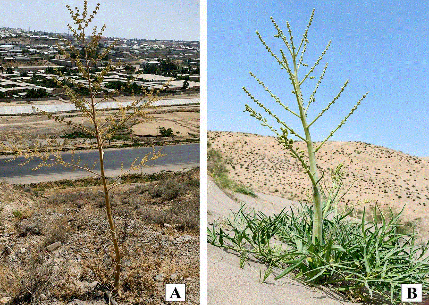
D. sabulosum is a rare endemic to Central Asia, considered endangered due to climatic stresses and overharvest for medicinal purposes (Pimenov, 2019). The conservation of these species is important not only for maintaining biodiversity but also for preserving potential of medicinal resources. However, traditional conservation methods may be insufficient due to the species’ specific reproductive strategies and vulnerable habitats. In vitro propagation techniques offer a promising approach for the conservation and sustainable utilization of rare and endangered plant species. The plants growing under natural conditions are shown in Figure 1.
To date, research has been conducted on microclonal propagation of D. ammoniacum D. Don, no research of the in vitro regeneration of D. microcarpum and D. sabulosum. This research presents a results of the biotechnological approaches for the conservation and potential commercialization of these two species.
The present study aims at establishing tissue culture protocols for plants regeneration through indirect somatic embryogenesis and indirect organogenesis for D. microcarpum and D. sabulosum. The effects of plant growth regulators on callus induction and subsequent plant regeneration, the use of different explant types were studied. This research is not only contribution to the conservation efforts but also the starting point for future studies on phytochemical properties and potential medicinal applications for these endangered species.
By developing of the efficient in vitro propagation methods, we aim to provide a sustainable approach to conserve and sustainable use of these species, as well as potential for new pharmacological researches and development of plant-based medicinal products.
Plant material (mature seeds) of D. microcarpum were collected in July 2022 from the Fergana Valley, Namangan, Davlatabad district, (Girvansai street) on Kokunbai road, (300–500 m above sea level, N 71 °599538; E 41 °01789). Seeds of the second species, D. sabulosum, were collected in July 2023 near the tugain-sandy reserve located in the middle reaches of the Amu Darya River, Bukhara region of the Republic of Uzbekistan (N 40°3402; E 62°1314). Identification of herbarium specimens collected during the study was carried out using Flora of Uzbekistan (Korovin, 1959) and Key to the Plants of Central Asia (Pimenov, 1983). Voucher specimens (Nos. 12072022 and 23062023) were deposited in the TASH Herbarium, Tashkent, Uzbekistan.
The experiment was conducted in the Biotechnology Laboratory of the Botanical Garden named after F.N. Rusanov at the Institute of Botany, Academy of Sciences of Uzbekistan, during 2022–2025.
Mature seeds of D. microcarpum were collected in July 2022 from the Fergana Valley, Namangan, Davlatabad district, (Girvansai street) on Kokunbai road, (300–500 m above sea level, N 71 °599538; E 41 °01789). Seeds of the second species, D. sabulosum, were collected in July 2023 near the tugain-sandy reserve located in the middle reaches of the Amu Darya River, Bukhara region of the Republic of Uzbekistan (N 40°3402; E 62°1314). The collected seeds were stored in closed paper bags at −10 °C and later transferred to a laboratory refrigerator for cold stratification at +4 °C for one month before processing. Seeds were treated with 70% ethanol for 1.5 min, then rinsed with sterile water for three times and then surface sterilized in 4% sodium hypochlorid solution for 20 min, followed by three washes with sterile water. The sterilized seeds were cultured on ј MS (Murashige and Skoog, 1962) medium supplemented with 30 g/l sucrose and 8 g/l agar. To overcome dormancy, the seeds were kept in a refrigerator (+4 °C) for 60 days. After cold stratification, the seeds were incubated at +23 ± 2 ° C under 16/8 h photoperiod provided by cool fluorescent lamps in culture room. The segments of the germinated seeds were used as a source of explants.
The followings were used as explants: mature zygotic embryos, segments of germinated seeds (roots, hypocotyl, and cotyledons); MS media with combinations of 2,4 D (0, 0.5, 1 mg/l), benzylaminopurine (0.2, 0.5, and 1 mg/l), indole 3-butyric acid (0.2, 0.5, and 1 mg/l); α-naphthaleneacetic acid (0.2, 0.5, and 1 mg/l); tidiazuron (0.2, 0.5, and 1 mg/l) and kinetin (0, 0.2, 0.5 or 1 mg/l) were tested. The pH of the medium was adjusted to 5.8, and growth regulators were added post-autoclaving. Germinated seeds (root, hypocotyl, and cotyledon segments) were cut by 0.5–1 cm in length. The explants were cultured in sterilized Petri dishes containing 25 ml of above-mentioned media combined with required doses of growth regulators and without them as a control.
Aseptically isolated mature zygotic embryos and the segments of the germinated seeds (root, hypocotyl, and cotyledon) were used as explants: root and hypocotyl were of 10 mm, cotyledon of 5 mm length. The 14–20 days-old seedlings were cut and the segments were cultivated on sterile MS medium with 2,4-D (0–0.5 mg/l), IBA (0–0.5 mg/l), in combination with TDZ (0.2, 0.5, and 1 mg/l) TDZ, Kin (0, 0.2, 0.5, and 1 mg/l), and BAP (0–1 mg/l) for callus induction. Calli were subcultured on the same medium every 4 weeks, each time divided into pieces of 2–4 mm diameter.
Mature zygotic embryos were placed on MS medium without a growth regulator to induce direct somatic embryogenesis. Cultures were incubated at +23 ± 2 ° C with 1,800 lux provided by cool fluorescent lamps with a photoperiod of 16/8 hours in culture room. Each experiment was performed with 5 replicates and the percentage of callus induction in the different media was recorded in eight weeks.
In two months after cultivation, the embryogenic callus was transferred to MS medium without growth regulators for induction of the embryogenesis. Mature somatic embryos were detected in 4 weeks after the incubation of the explants of nutrient media. The rate of indirect embryogenesis and mean number of embryos per explant was also calculated after 16 weeks. Then the mature somatic embryos were transferred to a hormone-free MS medium for germination and plantlet formation.
Morphogenic calli were transferred to the regeneration medium with various concentrations of BAP (0.1–1 mg/l), or combined with IBA (0–0.5 mg/l) or NAA (0–0.5 mg/l), to evaluate plants regeneration.
The share of explants with embryogenic callus was recorded in 4 weeks after culture. At this stage, the share of callogenesis, indirect somatic embryogenesis and number of embryos at different stages (globular, torpedo and cotyledonary), conversion of embryos into seedlings were also recorded.
Data analysis was displayed with box plots to reflect the distribution, mean, and standard deviation of measurements, conducted in triplicate (n = 3). Statistical analysis was done with one-way analysis of variance (ANOVA), followed by Tukey’s multiple comparison post hoc test with a 95% significance level (P < 0.05) for datasets that met normality assumptions. For datasets demonstrating non-normal distribution, exponential transformation was applied. When normalization was not achievable, the Kruskal-Wallis test, accompanied by Dunn’s test and Benjamini-Hochberg (BH) procedure, was applied. All statistical analyses were performed using RStudio, version 2023.06.1.
The initial experimental approach aimed to use mature zygotic embryos as explants for both D. microcarpum and D. sabulosum to expedite the process by bypassing seed germination. However, the two species showed markedly different responses, demanding species-specific adaptations in explant selection. For D. microcarpum, zygotic embryos showed no response, failing to induce callus or plant formation. This lack of reactivity prompted a shift in strategy, leading to the use of seedling segments derived from germinated seeds as the primary explant source for this species. In contrast, zygotic embryos proved to be functional explants for D. sabulosum, and the lake of in vitro germination of D. sabulosum seeds validated the continued use of its zygotic embryos as explants. The decision to employ distinct explants for each species was driven by these differential responses.
Mature zygotic embryos and the segments of the germinated seeds of two species D. microcarpum and D. sabulosum were used to induce callusogenesis. The callus formation was observed on root explants of D. microcarpum in 7 days after in vitro cultivation (Fig. 2A), in 15 days on hypocotyl segments of the germinated seeds, and no callus was observed on cotyledon segments. Callusogenesis was observed on the the zygotic embryos of D. sabulosum within 8 to 10 days. On hypocotyl segments callus primarily formed at the basal ends, whereas on roots callus was detected across the entire the surface as well as on zygotic embryos ( Figure 2). Root-derived callus was white, watery with a smooth surface, gradually becoming brown, whereas hypocotyl-derived callus was soft or friable, exhibiting a sticky consistency and nodular form with proembryogenic structures, ranging from yellow-white to creamy color ( Figure 3: 3B).
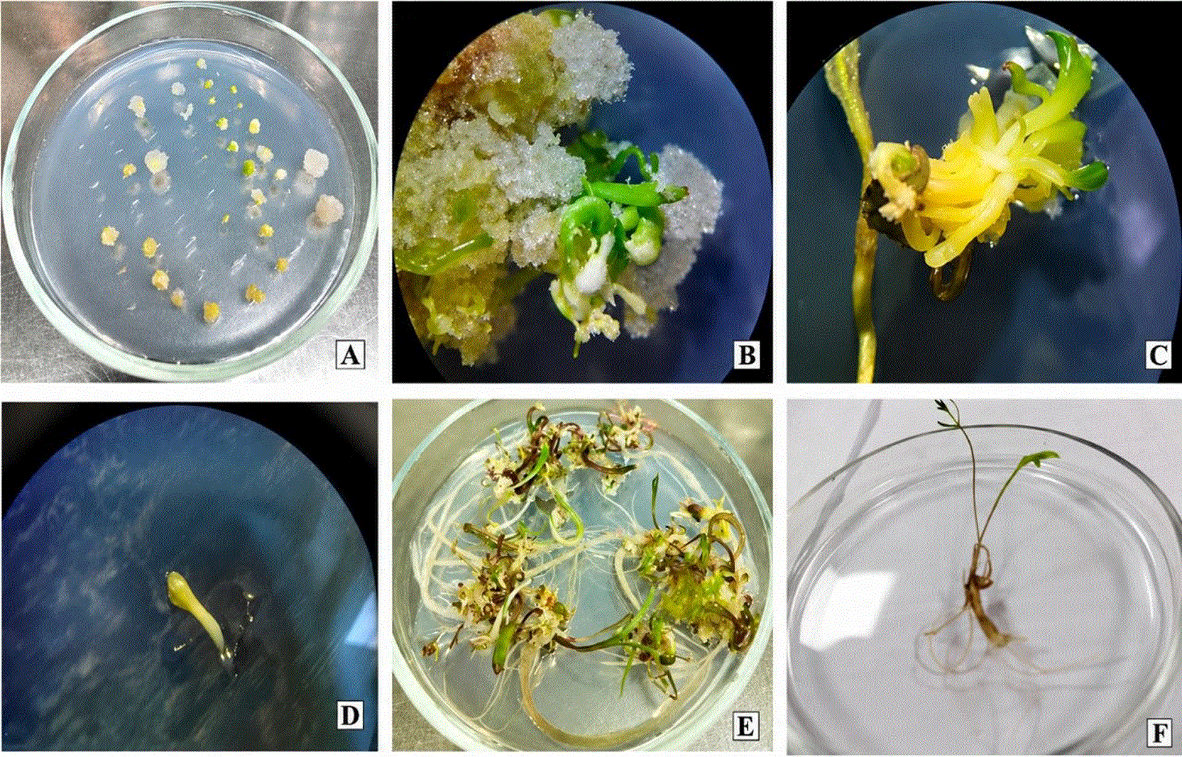
A. Callus induction on hypocotyls explants; B. induction of somatic embryos from hypocotyls explants; C. formation stage of somatic embryos in calli produced from hypocotyls explants on hormone free MS medium; D. isolated single somatic embryos, E.F. plant developed from somatic embryos.
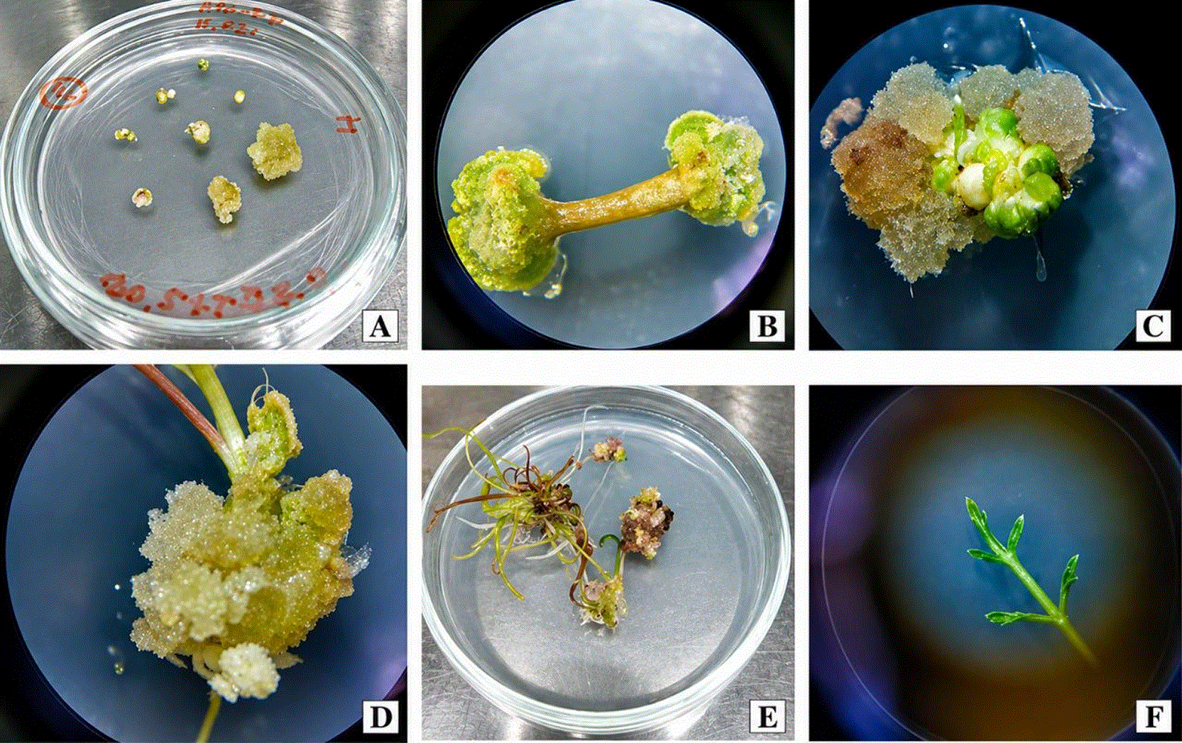
A. Callus induction in zygotic embryos explants; B. 2 weeks old compact callus obtained from zygotic embryos explants; C,D. initiation stage of shoot regeneration in calluses produced from zygotic embryos explants on MS medium supplemented 0.5 mg\l BAP and 0.5 mg\l IBA; E. shoot formation from the calli; F. plant regeneration.
The Kruskal-Wallis test results indicated significant differences in the impact of growth regulators on the callus induction in roots (χ2 = 51.13, p = 0.007) and hypocotyls (χ2 = 45.91, p = 0.024) in D. microcarpum. Further analysis using Dunn’s post-hoc test identified significant treatment effects in D. microcarpum compared to controls. For root explants, the application of 2,4-D (0.2 mg l−1) combined with TDZ (0.2 mg l−1) (93.3 ± 6.7%, Z = 3.47, p = 0.029) and combination of 2,4-D (0.5 mg l−1) + KIN (0.2 mg l−1) (93.3 ± 6.7%, Z = 3.47, p = 0.038) significantly increased callus induction over the control group ( Figure 3).
The Kruskal-Wallis test also revealed significant differences in callus induction across treatments in D. microcarpum hypocotyls (χ2 = 45.91, p = 0.0240). However, subsequent pairwise comparisons using Dunn’s test did not identify any specific growth regulator combinations that significantly differed from each other after correcting for multiple comparisons (Fig. 4. B). However maximum callus induction (73.3 ± 6.7%) was observed in treatments 2,4-D (0.2 mg l−1) + Kin (0.2 mg l−1) (73.3 ± 6.7%), 2,4-D (1.0 mg l−1) + Kin (0.2 mg l−1) (73.3 ± 13.3%), 2,4-D (0.5 mg l−1) + TDZ (0.2 mg l−1) (73.3 ± 6.7%) and 2,4-D (0.5 mg l−1) + TDZ (0.5 mg l−1) (73.3 ± 6.7%). In contrast, no significant differences were detected in the callus induction of D. sabulosum across different treatments (χ2 = 30.40, p = 0.3943). However, the maximum percentage of callus (93.3 ± 6.7%was observed in the treatment of 2,4-D (0.5 mg l−1) + TDZ (0.5 mg l−1) ( Figure 4).
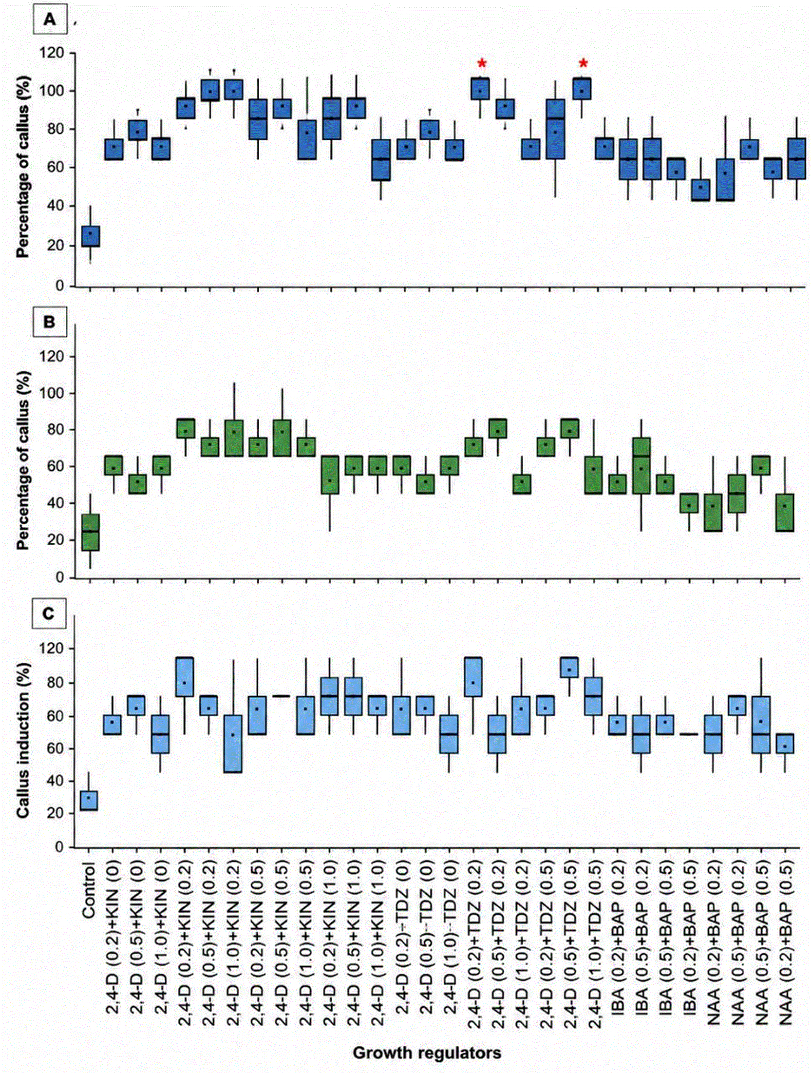
(A) Root explants of D. microcarpum. (B) Hypocotyl explants of D. microcarpum. (C) Callus induction in D.sabulosum. Asterisks (*) indicate significant differences identified by the Kruskal-Wallis test followed by Dunn’s post-hoc test (p < 0.05). The mean is represented by a dot (.) and the median by a horizontal line (—).
The study demonstrated significant variations in somatic embryogenesis responses across different growth regulator combinations in D. microcarpum. The most effective treatment, 2,4-D (0.5 mg l−1) combined with Kinetin (0.5 mg l−1), achieved the highest mean percentage of somatic embryogenesis at 66.67 ± 6.67% and the greatest average number of somatic embryos at 7.67 ± 0.33 per explant. 2,4-D (0.2 mg l−1) with Kin (0.5 mg l−1) and 2,4-D (0.2 mg l−1) with Kin (0.2 mg l−1), yielding average embryo numbers of 3.00 ± 1.00) and 3.33 ± 0.33) respectively ( Figure 5).
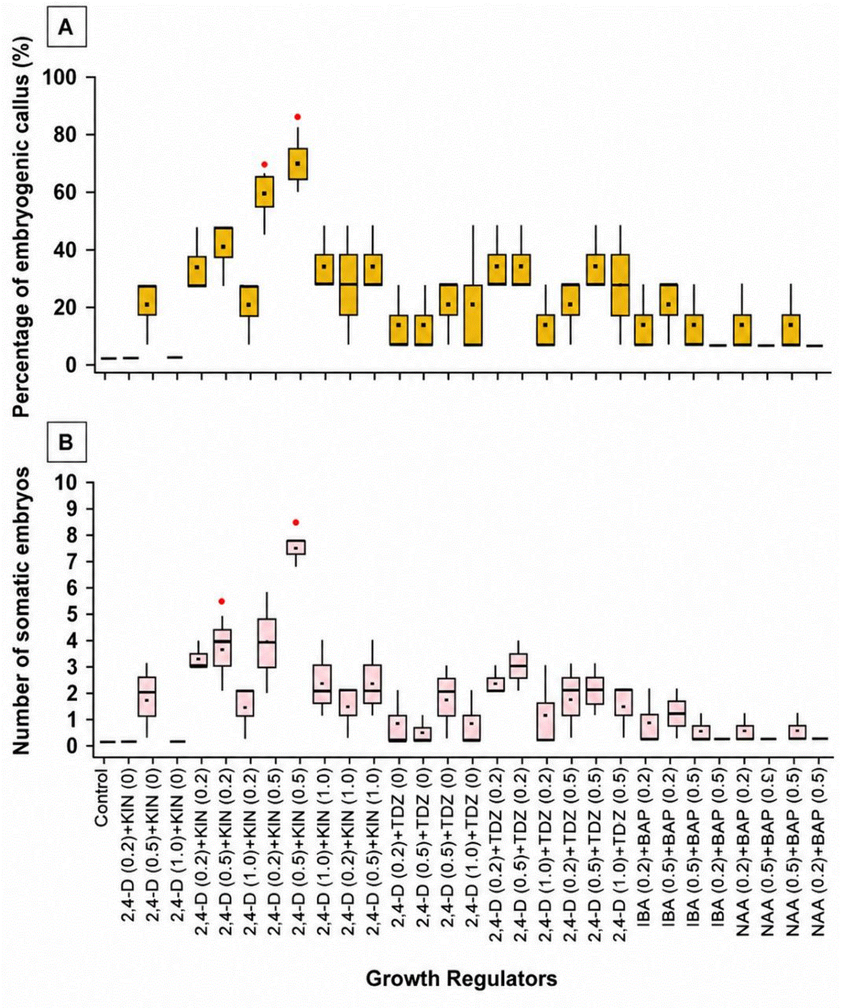
(A) Percentage of embryogenic callus. (B) Number of somatic embryos. Asterisks (*) indicate significant differences identified by the Kruskal-Wallis test followed by Dunn’s post-hoc test (p < 0.05). The mean is represented by a dot (.) and the median by a horizontal line (—).
Statistical analysis using the Kruskal-Wallis test confirmed significant differences both in embryogenic callus percentages (χ2 = 57.01, p = 0.0014) and somatic embryo numbers (χІ = 59.13, p < 0.0008) across treatments. Dunn’s post-hoc test further highlighted the superiority of 2,4-D (0.5 mg l−1) + Kin (0.5 mg l−1) over less effective treatments like 2,4-D (0.2 mg l−1) + KIN (0 mg l−1) and the control, with significant pairwise differences.
Upon transferring embryogenic calluses to hormone-free media in 10 weeks, microscopic examination revealed somatic embryos at various developmental stages, including globular, heart-shaped, torpedo, and cotyledonary forms. While most embryos exhibited normal morphology, some abnormalities occurred, such as embryos with multiple cotyledons, and sporadic secondary embryos developing on primary embryos.
D. sabulosum did not produce embryogenic calluses under any phytohormone combinations tested. Among the species analyzed, only D. microcarpum achieved a frequency of somatic embryogenesis ranging from 5% to 40%. After six weeks on a hormone-free medium, approximately 40–50% of cotyledonary somatic embryos germinated and developed into phenotypically consistent young plantlets, showcasing the potential for efficient clonal propagation and subsequent transplantation into pots for further growth and analysis ( Figure 2. C, D).
Following the production of primary somatic embryos, the embryogenic callus of D. microcarpum was transferred to MS medium supplemented with various concentrations of growth hormones to induce secondary somatic embryogenesis. One-way ANOVA revealed significant differences in the mean number of secondary somatic embryos among the different growth regulator treatments (p = 0.02). The control treatment produces the highest average number of secondary somatic embryos (15.33 ± 2.60) (Fig. 6). This was followed by 2,4-D (0.2 mg l−1) + Kin (0.5 mg l−1) and IBA (0.2 mg l−1) + BAP (0.5 mg l−1), both producing means of 11.67 ± 0.88 and 11.67 ± 1.45 embryos, respectively. In contrast, NAA (0.2 mg l−1) + BAP (0.5 mg l−1) resulted in the lowest production of secondary embryos (6.33 ± 0.33), significantly differing from the control ( Figure 6).
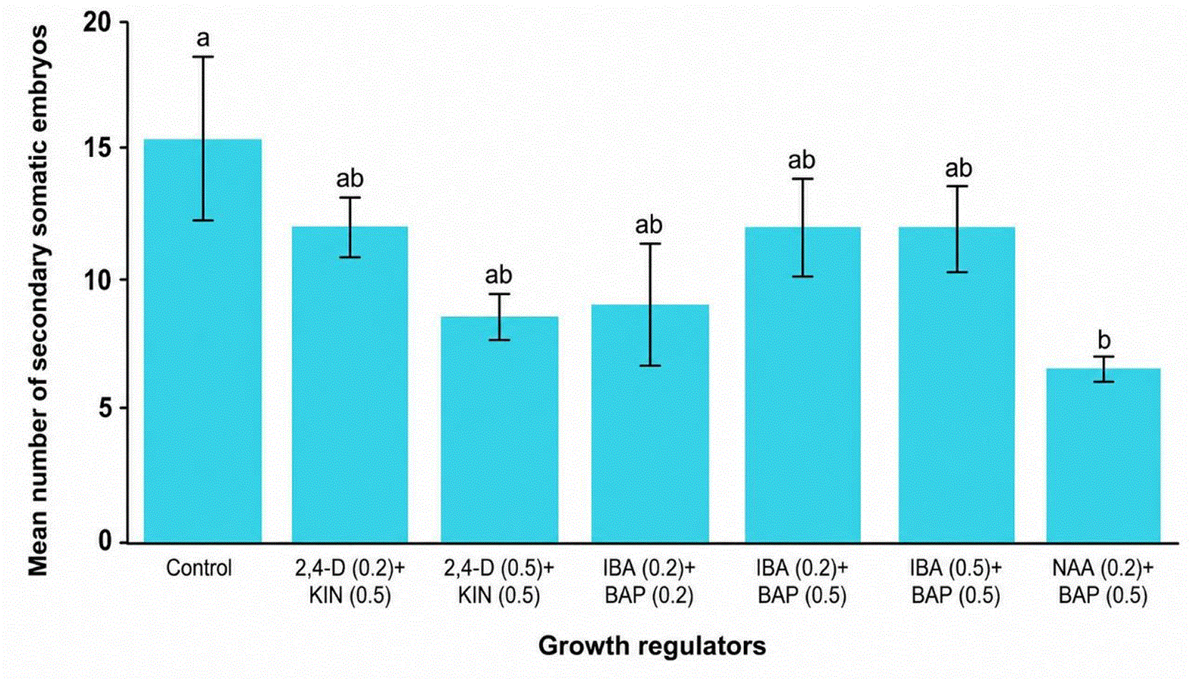
Data are expressed as mean ± standard error of the mean (SEM; n = 3). Different letters above the bars indicate statistically significant differences between treatments, as determined by Tukey’s test (p < 0.05).
The study evaluated the effectiveness of various growth regulator combinations on shoot and root formation in callus cultures of D. sabulosum using MS medium supplemented with different concentrations of BAP, NAA, and IBA. Calli obtained from zygotic embryos were quickly responsive, expanding and developing green spots within two weeks, which turned brown by the third week (Fig. 3 A and B). By the fifth week, green shoots began regenerating, with regeneration outcomes closely tied to the growth regulators used ( Figure 3. E and F).
The optimal shoot regeneration was observed with the combination of IBA (0.5 mg l−1) and BAP (0.5 mg l−1), achieving a regeneration rate of 66.7 ± 6.7% (Fig. 7. A). This was followed by IBA (0.2 mg l−1) with BAP (0.2 mg l−1), which resulted in a 53.3 ± 6.7% regeneration rate. Among BAP-only treatments, BAP (0.5 mg l−1) yielded 40 ± 11.6% shoot regeneration, while the control group showed a modest regeneration rate of 26.67 ± 6.67% ( Figure 7).
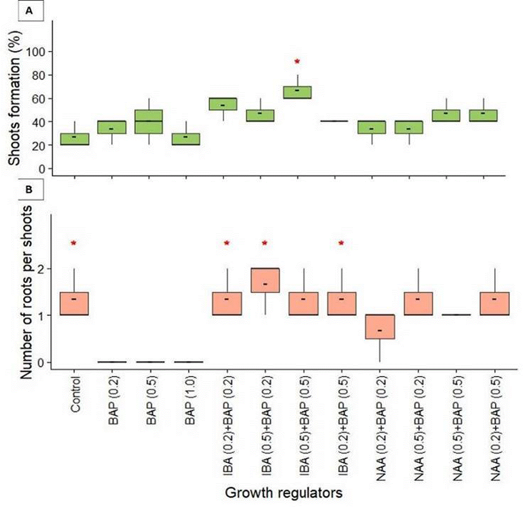
(A) Percentage of shoot formation. (B) Number of roots per shoot.
The Kruskal-Wallis test for shoot regeneration did not show significant overall differences among treatments (χ2 = 19.06, p = 0.060), but Dunn’s test revealed significant pairwise differences, notably between BAP (1.0 mg l−1) and IBA (0.5 mg l−1) + BAP (0.5 mg l−1) (Z = −3.06, p = 0.037), underscoring the influence of specific hormone combinations on regeneration outcomes.
For root formation, significant differences in the number of roots per shoot were observed (χ2 = 25.18, p = 0.0086). Control shoots produced an average of 1.3 ± 0.3 roots per shoot. Different concentrations of BAP alone (0.2, 0.5, and 1.0 mg l−1) inhibited root formation. This stark contrast with the control was statistically significant for all BAP concentrations (Dunn’s test, p < 0.05).
Combinations of hormones generally promote root formation. The IBA (0.5 mg l−1) + BAP (0.2 mg l−1) treatment yielded the highest mean number of roots per shoot (1.7 ± 0.3). Other IBA and BAP combinations, including IBA (0.2 mg l−1) + BAP (0.2 mg l−1), IBA (0.5 mg l−1) + BAP (0.5 mg l−1), and IBA (0.2 mg l−1) + BAP (0.5 mg l−1), all produced means equal to the control (1.3 ± 0.3 roots per shoot) ( Figure 7B). Post-hoc analysis revealed significant differences between BAP-only treatments and several hormone combinations. The most pronounced difference was observed between BAP (0.2) and IBA (0.5) + BAP (0.2) treatments (Z = −2.869428, p = 0.04523361). This comparison underscores the contrasting effects of BAP alone versus its combination with IBA on root formation.
Up to now, for two species of the genus Dorema (D.microcarpum and D.sabulosum) native to Uzbekistan there is no report on tissue culture for inducing callusogenesis somatic embryogenesis. In this study, we describe tissue culture protocols for the plantlet regeneration through indirect somatic embryogenesis and indirect organogenesis. Many studies on the microclonal propagation of species of the Apiaceae family use seedling segments sprouted from seeds as an explant. But in some studies (Bernard et al. 2007; Jamalova et al., 2022; Jamalova et al., 2025; Mustafina et al., 2025) and in our studies, zygotic embryos have also been used as an explant. The zygotic embryos of D. microcarpum did not give any results, in MS medium supplemented with different combinations of phytohormones prepared for the induction of callusogenesis or somatic embryogenesis was not changed. Therefore, we used seedling segments for this species.
In most combinations of phytohormones based on the MS nutrient medium, in which the successful development of the callus culture was observed, are present NAA and BAP (Zare et al., 2010; Sharifi et al., 1995; Tiwari et al., 2000; Pant et al., 2007; Mustafina et al., 2021, 2024). In the same study, Irvani et al. (2010) reported that BAP and NAA are important for the induction of callusogenesis in the D.ammoniacum. But in our studies, the use of low concentrations of 2,4-D and TDZ was successful for callusogenesis for all types of explants. The results of this experiment showed that 2,4-D (0.5 mg/L) + Kin (0.2 mg/L) followed by 2.4-D (0.5 mg/L) + Kin (0.5 mg/L) caused the highest somatic embryogenesis for hypocotyl explants of D. microcarpum. At that time, in D. sabulosum did not have somatic embryos in the explants of zygotic embryos and in the root explants of D. microcarpum too. Hyun-A Woo et al. (2021) reported that 2,4-D induced somatic embryogenesis. Mizukami et al. (2008) also reported the similar results. But in our studies, low concentrations of 2,4-D (0,2–0,5 mg/l) with Kinetin increased embryogenic calli. Explants planted in the medium of MS only with 2,4-D did not induce embryogenic calli. It was observed that in treatments with low concentration of 2,4-D without Kin, no embryo was observed. Therefore, we can conclude that Kin existence is very important for embryogenesis in D. microcarpum. Increase in the concentration of 2,4-D had a negative effect on somatic embryogenesis. This means that Kin is also necessary for the initiation of somatic embryogenesis. However, immature embryos did not form any embryogenic calli at all in the 1–2 mg/l 2,4-D treatments, even though the incubation period was prolonged. These results clearly showed that 1 mg/L 2,4-D is the most suitable concentration for embryogenic callus formation from hypocotyl explants. However, for the maturation of somatic embryos, 2,4-D must be removed from the nutrient medium. The same result is reported in the studies of Otroshy et al. (2013).
In this study, embryo maturation was not observed as long as the explants remained in the induction medium, and the origin of all stages of somatic embryogenesis occurs after the transition of explants to a hormone-free medium. Cho et al. (2003) reported that, in many plants such as carrot, after inducing in medium containing 2,4-D the somatic embryogenesis performed in hormone-free medium. The results of this research showed that the maturity of normal embryos was successful on hormone free MS medium as well. Our results showed that, the highest percentage of embryogenesis is related to hypocotyl explants. The first sign of somatic embryogenesis on embryogenic callus was marked by the appearance of globular structures that were attached to the surface by suspensor-like structures. Development of globular embryos appeared to progress through typical heart-, torpedo-shaped and cotyledonary embryos.
Indirect somatic embryogenesis was detected on hypocotyl segments of the germinated seeds of D. microcarpum, whereas callusogenesis was observed on root segments of the germinated seeds, and no processes were observed on the cotyledons. For D. sabulosum indirect organogenesis was observed on zygotic embryos used as explants. In this report, we describe for the first time a simple and effective protocols for micropropagation of D. microcarpum through somatic embryogenesis with the hypocotyl as the explants, and the protocol of micropropagation for D.sabulosum through indirect organogenesis with the zygotic embryos as explants.
However, further research is needed to determine cultivation conditions to increase the frequency of somatic embryogenesis induction and the rate of plant recovery from somatic embryos and acclimatization of regenerated plants ex vitro.
Not applicable. This systematic review did not involve any direct human or animal subjects, nor did it collect primary data requiring ethical approval.
All data generated or analyzed during this study are included in this published article.
This research was supported by the the projects “Digital Nature. Development of a digital platform for the flora of Central Uzbekistan,” implemented by the Institute of Botany of the Academy of Sciences of the Republic of Uzbekistan for the period 2025–2029, and the project Development of the Digital Platform of the Flora of the Ferghana Valley as a ‘Nature Imperilled Area’ (Project No. AL-9224104319). This research was also supported by the project titled “Assessing climate change adaptation in endangered plants of Uzbekistan: A DNA barcoding approach” (AL 9224104464).
The author(s) declared that no grants were involved in supporting this work.
© 2026 Jamalova D et al. This is an open access article distributed under the terms of the Creative Commons Attribution License, which permits unrestricted use, distribution, and reproduction in any medium, provided the original work is properly cited.
Current Reviewer Status:
AWAITING PEER REVIEW
AWAITING PEER REVIEW
?
Key to Reviewer Statuses VIEW HIDE
ApprovedThe paper is scientifically sound in its current form and only minor, if any, improvements are suggested
Approved with reservations A number of small changes, sometimes more significant revisions are required to address specific details and improve the papers academic merit.
Not approvedFundamental flaws in the paper seriously undermine the findings and conclusions