Просмотр новости
Найдите то, что Вас интересует
 Спутники
 РН
РН SpaceX
 Роскосмос
 NASA
NASA
СпутникиРНSpaceXРоскосмосNASABackground Uterine fibroids are the most common benign tumors in women, affecting up to 40–60% of women. Although frequently encountered during pregnancy, complications such as degeneration are uncommon. Conversely, small bowel obstruction (SBO) in pregnancy is a rare but potentially life-threatening condition, with an estimated incidence of 1 in 1,500 pregnancies. An association between SBO and uterine fibroids is exceptionally rare, with few cases reported in the literature. We present a unique case of SBO in pregnancy caused by a large degenerated fibroid, emphasizing the diagnostic and surgical challenges of this rare association. Case presentation A 31-year-old primigravida at 28 weeks of gestation presented with acute-onset pelvic pain and persistent vomiting. She was hemodynamically stable, afebrile, and had localized tenderness in the right iliac fossa without peritoneal signs. Fetal movements were active. Laboratory tests showed markedly elevated CRP. Abdominal ultrasound revealed jejunal distension, a large subserosal uterine fibroid with features of necrobiosis, moderate free intraperitoneal fluid, right-sided pyelocaliceal dilatation, and a gallbladder calculus. Initial management included nasogastric decompression and intravenous fluids. Due to persistent symptoms, an abdominopelvic CT scan showed small bowel dilatation with a transitional zone and a whirl sign around a twisted vascular pedicle adjacent to the fibroid, consistent with mechanical small bowel obstruction. Emergency laparotomy revealed a large subserosal fibroid with a small bowel loop adherent via an adhesive band forming a partial volvulus. No bowel ischemia was present. Adhesiolysis and division of the fibrous band relieved the obstruction without bowel resection. Postoperative recovery was uneventful, with reassuring fetal monitoring. At 35 weeks, she had spontaneous preterm labor and delivered a healthy male infant. Both mother and newborn remained well. Conclusion This case highlights the importance of timely diagnosis and intervention in mechanical bowel obstruction during pregnancy, ensuring favorable maternal and fetal outcomes despite preterm delivery.
Case Report
[version 1; peer review: awaiting peer review]
https://orcid.org/0000-0003-0034-7242
1, mehdi binous1, zeineb ben dhiafhttps://orcid.org/0009-0001-4818-8909
1, [...] Hamza Ben Abdallah1, Fahd Khefachahttps://orcid.org/0000-0003-0780-1671
1, mohamed raouf ben othman1, Maroua Yengui1, chiraz elfekih1https://orcid.org/0000-0003-0034-7242
1, mehdi binous1, [...] zeineb ben dhiafhttps://orcid.org/0009-0001-4818-8909
1, Hamza Ben Abdallah1, Fahd Khefachahttps://orcid.org/0000-0003-0780-1671
1, mohamed raouf ben othman1, Maroua Yengui1, chiraz elfekih11 University of Tunis El Manar Faculty of Medicine of Tunis, Tunis, Tunis, Tunisia
Oumayma Mejri
Roles: Conceptualization, Supervision, Validation, Writing – Review & Editing
mehdi binous
Roles: Conceptualization, Writing – Review & Editing
zeineb ben dhiaf
Roles: Resources, Validation
Hamza Ben Abdallah
Roles: Writing – Original Draft Preparation
Fahd Khefacha
Roles: Writing – Review & Editing
mohamed raouf ben othman
Roles: Data Curation, Investigation
Maroua Yengui
Roles: Writing – Review & Editing
chiraz elfekih
Roles: Validation
OPEN PEER REVIEW
REVIEWER STATUS AWAITING PEER REVIEW
Corresponding authors: Oumayma Mejri, mehdi binous Competing interests: No competing interests were disclosed.
Grant information: The author(s) declared that no grants were involved in supporting this work.
Copyright: © 2026 Mejri O et al. This is an open access article distributed under the terms of the Creative Commons Attribution License, which permits unrestricted use, distribution, and reproduction in any medium, provided the original work is properly cited. How to cite: Mejri O, binous m, ben dhiaf z et al. Case Report: Intestinal Obstruction in Pregnancy Due to a Degenerating Uterine Leiomyoma [version 1; peer review: awaiting peer review]. F1000Research 2026, 15:1274 (https://doi.org/10.12688/f1000research.180014.1) First published: 01 Aug 2026, 15:1274 (https://doi.org/10.12688/f1000research.180014.1) Latest published: 01 Aug 2026, 15:1274 (https://doi.org/10.12688/f1000research.180014.1)
Uterine fibroids are the most common benign tumors in women, affecting up to 40–60% of women of reproductive age. Although frequently encountered during pregnancy, they rarely cause complications such as degeneration or acute abdominal events. Small bowel obstruction (SBO) in pregnancy is uncommon, with an estimated incidence of 1 in 1,500, but it carries significant maternal and fetal risks. The occurrence of SBO caused by uterine fibroids is exceptionally rare, with few cases reported. Here, we present a case of SBO in pregnancy due to a large degenerated fibroid, illustrating the diagnostic and surgical challenges of this unusual presentation.
A 31-year-old primigravida, blood group O Rh-positive, with no significant past medical or surgical history, was admitted at 28 weeks of gestation with acute-onset pelvic pain and persistent vomiting.
On initial clinical evaluation, the patient appeared in good general condition, with stable hemodynamic and respiratory parameters and was afebrile. Abdominal examination revealed a distended but soft abdomen, with localized tenderness over the right iliac fossa and no signs of peritoneal irritation. Fetal movements were active and regular. There were no uterine contractions, vaginal bleeding, nor abnormal vaginal discharge.
Laboratory investigations revealed a hemoglobin level of 10.9 g/dL and a white blood cell count of 9,000/mm3. C-reactive protein (CRP) was markedly elevated at 650 mg/L. Renal function was within normal limits, as were serum electrolytes and lipase levels.
Abdominal ultrasound demonstrated marked fluid-filled distension of the jejunal loops, with a maximal diameter of 30 mm. The intestinal loops appeared displaced by a large subserosal uterine fibroid showing features of necrobiosis, extending superiorly to the left subphrenic region. A moderate amount of free intraperitoneal fluid was noted in the subhepatic space and between the bowel loops. Additionally, a simple gallbladder calculus was observed.
Given the patient’s persistent vomiting and the ultrasound findings of marked small bowel distension, a diagnosis of intestinal obstruction was considered.
A surgical opinion was requested. The patient was initially managed conservatively with nasogastric decompression, intravenous fluids, and close hemodynamic monitoring. Due to the persistence of symptoms and diagnostic uncertainty, an abdominopelvic CT scan was performed.
CT imaging revealed small bowel dilatation, with the maximal diameter measuring 35 mm. A transitional zone was observed, characterized by a whirl sign, indicating a volvulus, centered around a twisted vascular pedicle adjacent to a large subserosal fibroid (Figure 1). The fibroid measured 11 × 18 cm, located posterolaterally on the left side of the uterus, with spontaneous hypodensity, heterogeneous enhancement, and extensive necrosis (Figures 2, 3). It displaced adjacent bowel loops toward the right side. There was associated circumferential thickening of the involved bowel wall, no evidence of pneumoperitoneum, and moderate intra-abdominal fluid. These findings were consistent with acute mechanical small bowel obstruction, with the transitional point located in the supramesocolic region.
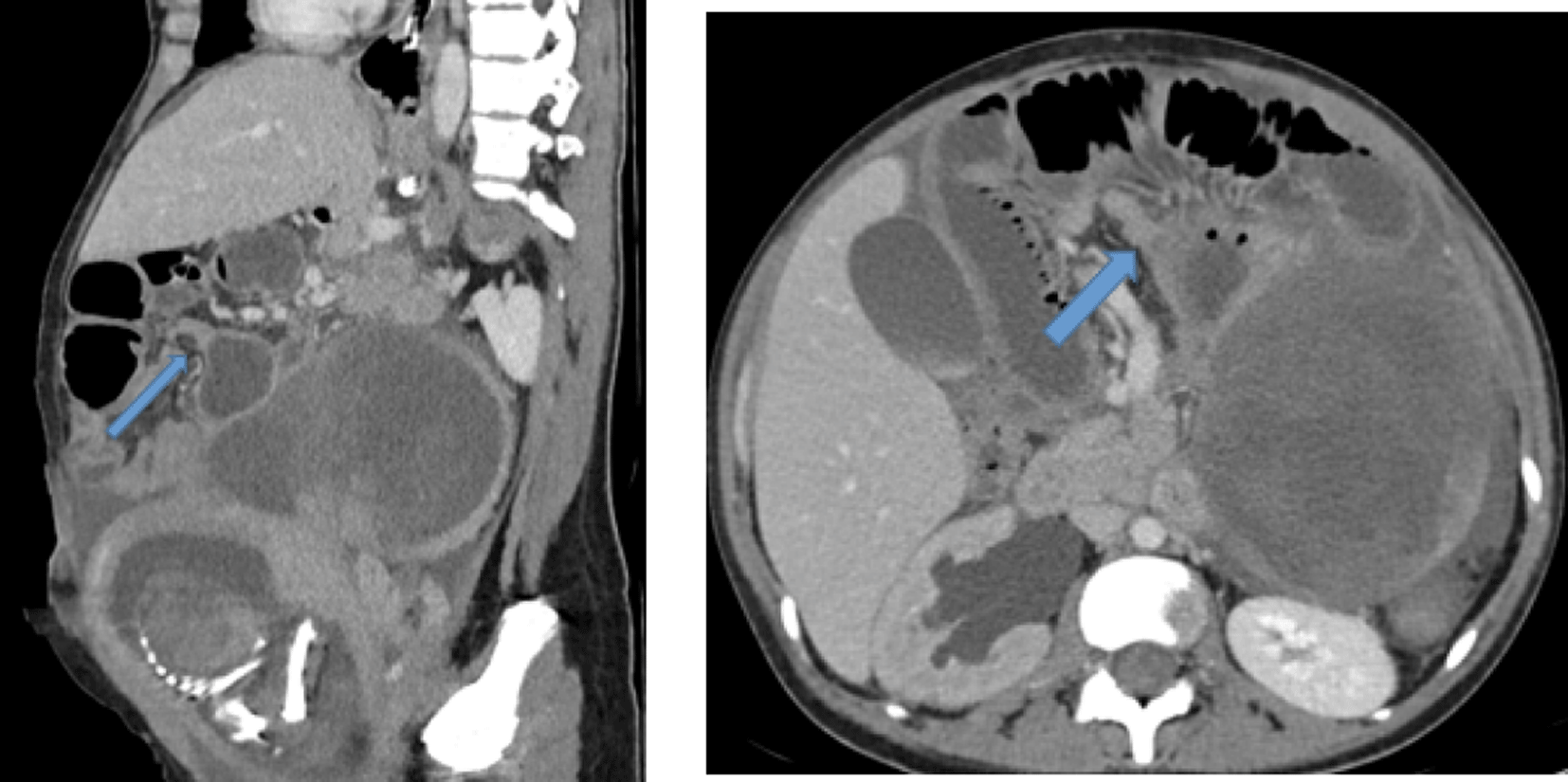
Axial and sagittal CT scans showing dilatation of the jejunal loops upstream of a supramesocolic transition zone (blue arrow).
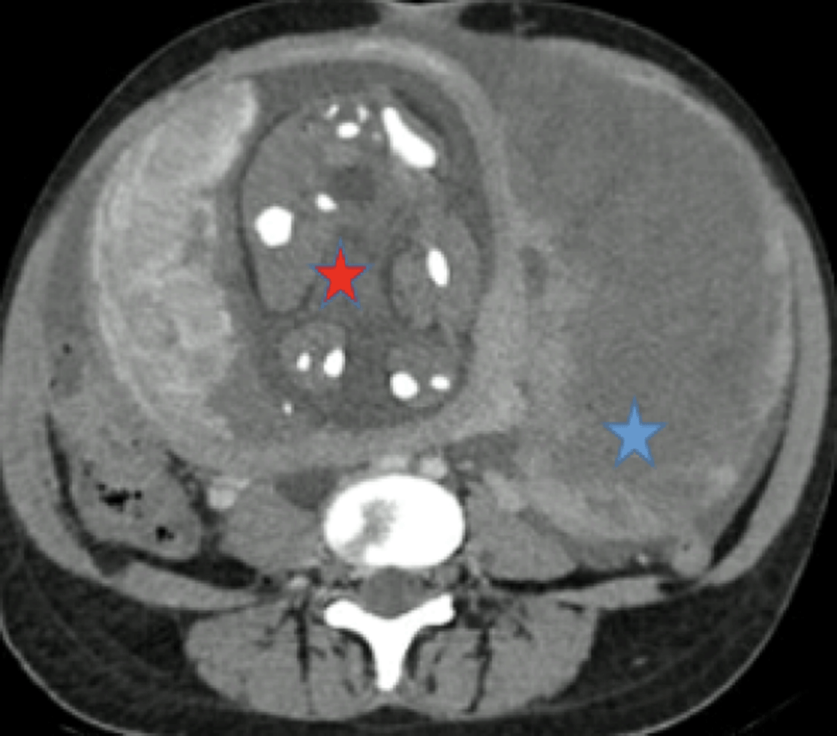
Axial CT slice showing the fetus (red star) and a left-lateralized fibroid with central necrosis (blue star).
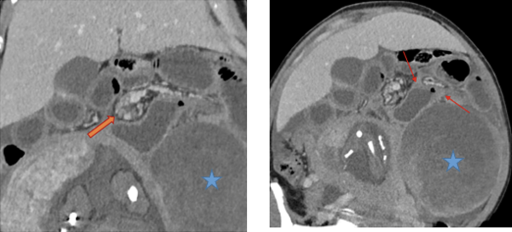
Abdominal CT showing a double-beak sign (orange arrow) suggestive of a closed-loop obstruction, with a characteristic “whirl sign” (red arrows) adjacent to the fibroid (blue star).
The final working diagnosis was a mechanical intestinal obstruction due to strangulation.
The patient was transferred to the surgical unit and underwent emergency infraumbilical midline laparotomy.
During surgery, a small amount of intraperitoneal fluid was noted. A large subserosal uterine fibroid was identified (Figure 4), occupying the left abdominal quadrant. A loop of small bowel, approximately 1.5 meters from the ileocecal junction, was found adherent to the fibroid, forming a partial volvulus around its mesentery, without signs of ischemia. Additionally, a loose fibrous band was found between a small bowel loop and the round ligament. The bowel appeared distended upstream but viable ( Figure 5). Thus, surgical exploration revised the initial diagnosis of volvulus and revealed that the intestinal obstruction was in fact caused by an adhesive band.
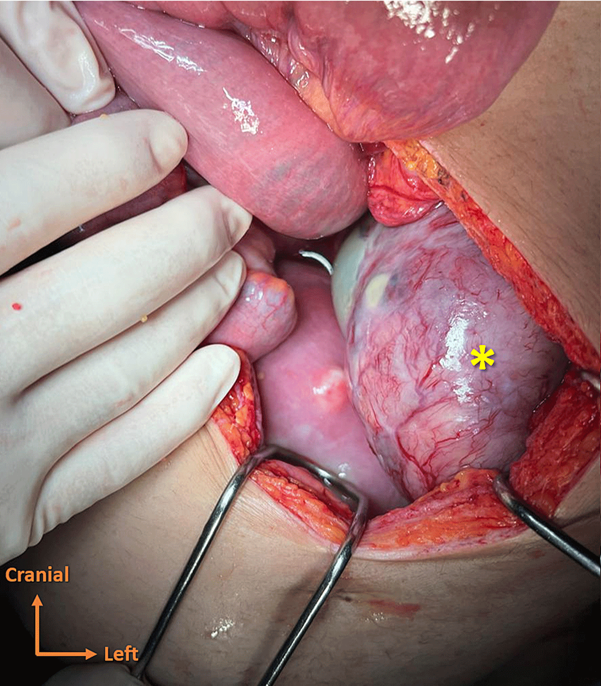
Perioperative view showing a large subserosal uterine fibroid identified (yellow asterisk).
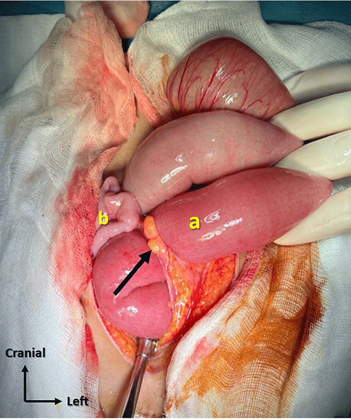
Perioperative view showing an adhesion between a small bowel loop and the round ligament (black arrow). Dilated small bowel loops (a) contrast with collapsed loops (b), with partial reduction of the small bowel incarceration.
Surgical management included careful release of the adhesion between the bowel loop and the fibroid, division of the fibrous band, and retrograde decompression of the bowel. No bowel resection was necessary. Hemostasis was secured, and the abdomen was closed.
Her recovery was uneventful, with normalization of inflammatory markers and improvement of clinical symptoms. She received tocolysis, prophylactic low-molecular-weight heparin (LMWH), and appropriate analgesia. Obstetrical monitoring was reassuring, and the pregnancy was allowed to continue.
At 35 weeks of gestation, the patient presented with pelvic pain suggestive of uterine contractions. She subsequently underwent a spontaneous vaginal delivery of a healthy male infant weighing 2,275 grams, with Apgar scores of 9, 10, and 10 at 1, 5, and 10 minutes, respectively. The postpartum period was uneventful, and both mother and newborn remained in good condition. She was discharged home with scheduled outpatient follow-up.
Uterine fibroids, or leiomyomas, are the most common benign tumors in women of reproductive age, with an estimated prevalence of 20–40%.1,2 During pregnancy, fibroids are present in approximately 1–4% of cases and may be associated with various complications depending on their size, number, and location.2,3 The most frequent obstetric complications include pain due to degeneration (necrobiosis), miscarriage, preterm labor, placental abruption, malpresentation, and postpartum hemorrhage.3
Among these, aseptic necrobiosis is a relatively common complication during pregnancy, particularly in the second and third trimesters. It occurs when fibroid needs outpaces its blood supply, leading to ischemia and tissue degeneration. Clinically, necrobiosis typically presents with acute localized abdominal or pelvic pain, low-grade fever, leukocytosis, and elevated inflammatory markers.4,5 The diagnosis is primarily clinical, supported by imaging findings. Ultrasound may show heterogeneous echogenicity within the fibroid, but MRI is considered more sensitive.6
In contrast, acute intestinal obstruction (AIO) during pregnancy is a rare but serious surgical emergency, with an estimated incidence of 1 in 1,500 to 66,000 pregnancies.7 The most common causes include adhesions (58%), volvulus (24%), hernias (8%), and less commonly, neoplasms, intussusception, or Crohn’s disease.8 Obstruction due to extrinsic compression by a uterine fibroid is exceedingly rare, and only isolated case reports have been described in the literature.9
In the present case, the subtle clinical presentation and the lack of complete bowel movement or flatus cessation rendered the diagnosis of intestinal obstruction difficult. The challenge was further heightened by pregnancy-related anatomical changes, as the enlarging uterus displaces abdominal organs and can obscure typical clinical signs.10
Ultrasound was useful as an initial imaging modality, but due to diagnostic uncertainty, a contrast-enhanced CT scan was performed, which revealed classic signs of mechanical small bowel obstruction (dilated proximal loops, a transition point, and the whirl sign). Although CT imaging is generally avoided during pregnancy, it remains justified in urgent scenarios where maternal risk outweighs potential fetal radiation exposure. In such situations, the second and third trimesters are relatively safer, and efforts should be made to minimize dose and use shielding.11
Surgical exploration confirmed the obstruction of the small bowel loop caused by adhesion between a necrotic fibroid and an adjacent bowel loop, along with a fibrous band. While volvulus is a known cause of AIO in pregnancy, its association with fibroid-induced adhesions or compression is exceedingly rare. Only a few cases of small bowel obstruction secondary to giant subserosal fibroids have been reported.5,12 In these cases, the fibroid either compresses the bowel or forms adhesions that lead to torsion and obstruction.
In fact, fibroid necrobiosis leads to the release of cellular debris and inflammatory mediators. This local reaction causes peritoneal irritation and a fibrin-rich exudate. Progressive organization of this fibrin results in fibrous adhesions between pelvic organs. Thus, adhesions arise from peritoneal inflammation secondary to myometrial necrosis.5
Importantly, the absence of bowel ischemia or necrosis in this case allowed for a conservative surgical approach, with simple adhesiolysis and decompression. Avoiding bowel resectionreduced surgical morbidity and preserved maternal-fetal outcomes.
Early diagnosis of strangulated intestinal obstruction is crucial, as it significantly improves prognosis by allowing timely surgical intervention. In our case, the prompt recognition and management of the obstruction contributed to an uneventful maternal recovery and the continuation of pregnancy to late preterm (35 weeks) without complications.
Although our patient’s maternal recovery was uneventful, she did ultimately deliver preterm. This spontaneous preterm birth may be explained by the large fibroid and its complications. Recent work proposes that fibroid necrosis and the associated inflammatory reaction (release of cytokines, prostaglandins) may trigger uterine contractility and preterm labour.5,13
This case highlights the importance of considering non-obstetric causes of abdominal pain and vomiting in pregnancy, particularly when initial conservative management fails or when imaging suggests gastrointestinal involvement. Multidisciplinary collaboration, involving obstetricians, surgeons, and radiologists, is essential to ensure accurate diagnosis and optimal outcomes for both mother and fetus.This case has some limitations. First, although the intraoperative findings strongly suggested that inflammation secondary to fibroid degeneration promoted adhesion formation, the exact pathophysiological mechanism cannot be definitively established from a single case. Second, because of the exceptional rarity of this presentation, our findings cannot be generalized and should instead be interpreted as evidence that raises awareness of this uncommon cause of intestinal obstruction during pregnancy.
Written informed consent for publication of the patient’s clinical information and accompanying images was obtained from the patient. Copies of the signed informed consent forms in both Arabic and French have been submitted to the journal as supporting documentation. All reasonable efforts have been made to protect the patient’s anonymity; however, complete anonymity cannot be guaranteed.
ChatGPT, OpenAI was used solely as a language and writing assistance tool to improve the clarity, grammar, style, and organization of the manuscript. The AI tool was not used to generate, analyze, interpret, or modify any clinical data, results, images, or scientific conclusions. All clinical information, data collection, analysis, interpretation, and final manuscript content were reviewed and validated by the authors, who take full responsibility for the accuracy and integrity of the work.
No datasets were generated or analyzed during the current study.
The authors would like to thank all healthcare professionals involved in the patient’s care and management.
The author(s) declared that no grants were involved in supporting this work.
© 2026 Mejri O et al. This is an open access article distributed under the terms of the Creative Commons Attribution License, which permits unrestricted use, distribution, and reproduction in any medium, provided the original work is properly cited.
Current Reviewer Status:
AWAITING PEER REVIEW
AWAITING PEER REVIEW
?
Key to Reviewer Statuses VIEW HIDE
ApprovedThe paper is scientifically sound in its current form and only minor, if any, improvements are suggested
Approved with reservations A number of small changes, sometimes more significant revisions are required to address specific details and improve the papers academic merit.
Not approvedFundamental flaws in the paper seriously undermine the findings and conclusions