Просмотр новости
Найдите то, что Вас интересует
 Спутники
 РН
РН SpaceX
 Роскосмос
 NASA
NASA
СпутникиРНSpaceXРоскосмосNASAAbstract* Introduction Brucellosis is a zoonotic infection with diverse clinical manifestations. Cervical involvement is exceptionally rare and can resemble acute bacterial cellulitis. We report a case of brucellosis presenting as acute cervical cellulitis, along with a review of the literature. Case presentation We report the case of a 67-year-old man presenting with febrile cervical swelling. Evaluation included clinical examination, cervical CT scan, bacteriological analysis of abscess pus, and brucellosis serology. CT imaging showed a right latero-cervical collection. Culture of abscess pus isolated Brucella Melitensis and serology was positive. The patient underwent surgical drainage combined with targeted antibiotic therapy, with favorable outcome. Conclusion Cervical brucellosis is a rare presentation that may mimic common bacterial neck infections. Early diagnosis and appropriate management are essential for favorable outcome.
Brucellosis is one of the most common bacterial zoonoses worldwide and remains particularly endemic in countries of the Mediterranean basin.1 In Tunisia, it continues to be encountered, facilitated by traditional livestock farming practices and increased contamination of herds that escape vaccination, thus constituting a persistent public health problem.2 The clinical manifestations of brucellosis are generally nonspecific and may involve multiple organs. While osteoarticular, hepatosplenic, and neurological involvements are well described, soft tissue localization, especially in the cervical region, remains exceptional and is rarely reported in the literature. Cervical cellulitis of brucellar origin therefore represents a rare and atypical presentation of the disease, potentially mimicking other more common infectious etiologies.
We report a case of a patient presenting with acute cervical cellulitis and abscess caused by Brucella melitensis, confirmed microbiologically. Diagnosis was challenging due to nonspecific symptoms. Microbiological confirmation from abscess pus underscores the uniqueness of this case.
The patient was a 67-year-old male with no significant medical history, living in a rural area and having close and regular contact with cattle. He is an active smoker with an estimated 15 pack-years and reports regular consumption of unpasteurized milk.
He presented to our emergency department with a painful right-sided cervical swelling associated with fever and general deterioration, evolving over seven days.
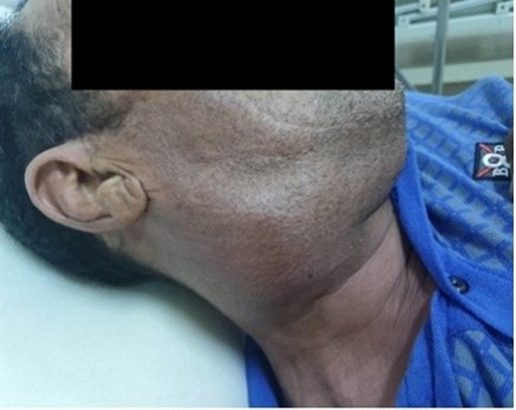
On clinical examination, a right latero-cervical mass measuring approximately 7 cm in its greatest diameter was noted. The mass was firm and tender on palpation, with overlying inflammatory skin changes ( Figure 1). Oropharyngeal examination and nasofibroscopy were unremarkable. The remainder of the physical examination revealed no significant abnormalities.
Initial laboratory investigations revealed a biological inflammatory syndrome, with a leukocyte count of 9,060 cells/mm3 and a marked elevation of C-reactive protein (CRP) to 142 mg/L.
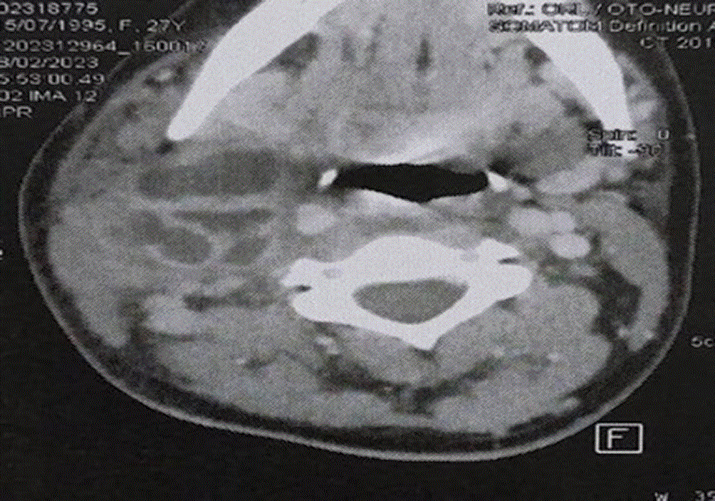
Cervicothoracic computed tomography demonstrated a right cervical collection measuring 38 × 78 mm, heterogeneous in appearance, with hypodense content and surrounded by a contrast-enhancing wall. There was also fat stranding involving the parotid, sublingual, parapharyngeal, and retropharyngeal spaces, associated with myositis of the right sternocleidomastoid muscle ( Figure 2). No osteoarticular or pulmonary lesions were identified.
Aspiration of the collection with pus sampling was performed, followed by bacteriological analysis. Culture allowed the isolation of Brucella melitensis. Brucellosis serology was positive, demonstrating the presence of specific IgM and IgG antibodies.
Emergency surgical drainage of the cervical abscess was carried out, combined with initial empiric antibiotic therapy. After etiological confirmation by culture and serological results, treatment was adjusted to a specific antibiotic regimen consisting of doxycycline at a dose of 200 mg per day and rifampicin at a dose of 600 mg twice daily, administered orally for six weeks.
The case was reported to the health authorities (brucellosis is a notifiable disease in Tunisia).
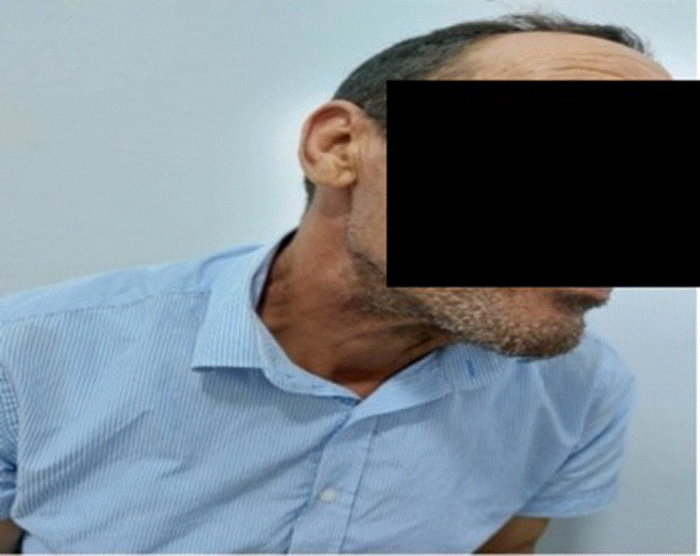
The outcome was favorable, with marked clinical improvement ( Figure 3) and progressive normalization of biological parameters. Follow-up cervical ultrasonography performed at the end of treatment showed no residual abnormalities.
Brucellosis is a worldwide zoonotic bacterial infection primarily caused by Brucella melitensis and Brucella abortus. It remains endemic in the Mediterranean basin, the Middle East, parts of Asia, and Latin America, with evidence of re-emergence in regions characterized by intense livestock activity.3,4 In Tunisia, the disease persists in an endemic-epidemic pattern, with heterogeneous and sometimes misleading clinical presentations.2 Recent meta-analyses highlight emerging antimicrobial resistance patterns in Mediterranean countries, raising increasing therapeutic concerns.1 In China, epidemiological data show rising incidence in certain rural provinces, largely associated with intensive animal farming and livestock movement.5,6
Transmission to humans occurs primarily from infected animals and most frequently through ingestion of unpasteurized dairy products. Transmission may also occur through direct contact with infected animals or their secretions during farming, slaughtering, or veterinary activities via breaches in the skin or mucosal surfaces. Less commonly, infection may result from inhalation of contaminated aerosols, particularly in laboratories or abattoirs.7 Human-to-human transmission remains exceptional. Epidemiological analyses demonstrate that rural settings and areas with insufficient veterinary control are at highest risk.5,6
From a pathophysiological perspective, Brucella is a facultative intracellular Gram-negative coccobacillus with a particular tropism for the reticuloendothelial system. After entry into the host, the bacteria are phagocytosed by macrophages, where they survive and replicate by inhibiting phagosome–lysosome fusion. This immune evasion capacity explains the subacute or chronic course of the disease, frequent relapses, and focal organ involvement.3,7 Soft tissue localization may result from hematogenous dissemination during bacteremia, extension from infected lymph nodes, or, more rarely, direct inoculation, mainly in occupational settings. Among the species of the genus Brucella, Brucella melitensis is the most frequently implicated in human infection and is considered the most pathogenic.5
In humans, brucellosis is clinically classified according to the duration of symptoms into three forms: acute when the disease evolves for less than eight weeks, subacute when symptoms persist between eight and fifty-two weeks, and chronic when the disease lasts for more than one year.8
Clinically, human brucellosis typically presents with undulating fever, profuse night sweats, arthralgia, fatigue, and hepatosplenomegaly. Osteoarticular complications are the most common, particularly sacroiliitis and spondylodiscitis. Neurological, cardiac, or genitourinary involvement may occur in complicated cases.7,9 Large clinical series emphasize the marked heterogeneity of manifestations, making purely clinical diagnosis unreliable.6,9 Soft tissue involvement in the form of cellulitis or abscess remains exceptional and represents an atypical presentation rarely described in the literature. Cervical soft tissue brucellosis may clinically mimic common bacterial deep neck infections. However, certain elements should raise suspicion in endemic areas, including a subacute course, epidemiological exposure such as livestock contact or consumption of unpasteurized dairy products, moderate leukocytosis despite markedly elevated inflammatory markers, and the presence of associated systemic symptoms such as fever, night sweats, and asthenia. Radiological findings on computed tomography or magnetic resonance imaging are nonspecific and typically demonstrate a rim-enhancing collection with surrounding inflammatory changes.10,11
Biological diagnosis relies on a combination of hematological, serological, and microbiological findings. Laboratory abnormalities may include mild anemia, leukopenia, thrombocytopenia, and moderate elevation of liver enzymes.12 Standard agglutination tests (e.g., Wright, Coombs) demonstrate good sensitivity and specificity when interpreted in the appropriate clinical and epidemiological context13 . Blood culture remains the gold standard but requires specialized techniques and prolonged incubation.14 Molecular methods improve early detection but are not consistently available in endemic areas.14
Therapeutic management is based on prolonged combined antibiotic therapy to ensure complete eradication of the pathogen and to prevent relapse, which is frequent in cases of insufficient treatment. Outside complicated forms such as endocarditis, spondylodiscitis, or neurobrucellosis, treatment relies on one of three recommended regimens combining two antibiotics active against Brucella for a minimum duration of six weeks.1 The first regimen combines doxycycline at a dose of 200 mg per day for six weeks with streptomycin at a dose of 1 g per day administered intramuscularly or intravenously during the first two to three weeks. The second regimen combines doxycycline with gentamicin at a dose of 3–5 mg/kg per day administered intramuscularly or intravenously for one to two weeks. The third regimen consists of doxycycline combined with rifampicin at a dose of 600–900 mg per day for six weeks.15 Surgical management is indicated in specific situations, including abscess formation, infected prostheses, compressive inflammatory masses, or endocarditis associated with large vegetations. Despite appropriate management, relapse rates remain significant, ranging from 5% to 20% according to published series.15
Cervical cellulitis represents a rare and atypical manifestation of brucellosis, which may be confused with more common bacterial soft tissue infections. This observation highlights the importance of considering brucellosis in the differential diagnosis of any unusual cervical cellulitis, particularly in endemic areas.
Written informed consent was obtained from the patient for publication of this case report and accompanying images.