Просмотр новости
Найдите то, что Вас интересует
 Спутники
 РН
РН SpaceX
 Роскосмос
 NASA
NASA
СпутникиРНSpaceXРоскосмосNASABackground Scoring of estrogen receptor (ER) and progesterone receptor (PR) expression in breast cancer is critical for identifying patients who would benefit with hormonal therapy. Since manual scoring of immunohistochemistry (IHC) is influenced by pathologist experience, fatigue, inter-observer variability, and subjectivity, artificial intelligence (AI)–based algorithms, trained on large datasets can aid to improve diagnostic accuracy. Methodology This study evaluated an AI-based algorithm for ER and PR IHC scoring in 297 ER and 293 PR cases of invasive breast carcinoma and compared the scores with that of pathologists (two senior and two junior) A pre-trained automated algorithm (Mimansa) identified region of interest and provided the scoreswhich was compared with the consensus score of pathologists-ground truth(GT).Concordance was evaluated using Cohen’s kappa and F1 score. Results For ER IHC, GT scores included 169 strong positive, 31 low positive, and 98 negative cases. Agreement with GT was 99% and 98% for senior pathologists, 97% for the AI algorithm, and 95% and 93% for junior pathologists. The algorithm correctly classified all strong positive cases but showed discordance in 16 low-score cases, with four false negatives and ten false positives. Notably, it identified two true positive cases missed by all pathologists. For PR IHC, agreement rates were 98% and 97% for senior pathologists, 92% for the algorithm, and 93% and 91% for junior pathologists. The algorithm achieved perfect accuracy in strong positive cases but produced 16 false negatives and eight false positives among low-score cases. Cohen’s kappa values were 0.91 (ER) and 0.84 (PR). Conclusion: The AI algorithm demonstrated high concordance with expert consensus, performing comparably to senior pathologists and outperforming junior pathologists in several metrics. It shows promise as a supportive second-reader tool, particularly in low-positive cases where diagnostic errors may significantly impact patient management.

Pai K, Singh BMK, Udupa CB et al. Leveraging Artificial Intelligence (AI) Based Algorithm for Accurate Estrogen Receptor (ER) and Progesterone Receptor (PR) Analysis in Breast Cancer Diagnostics: Potential to be a Crucial Aid in Routine Workflow [version 2; peer review: 2 approved]. F1000Research 2026, 15:702 (https://doi.org/10.12688/f1000research.175934.2)
Research Article
Revised
[version 2; peer review: 2 approved]
https://orcid.org/0000-0002-6947-5552
1, Brij Mohan Kumar Singh1, Chethana Babu Udupa1, [...] Madhavi Pai1, Vani Verma1, Sumit Jha2, Kiran Aatrehttps://orcid.org/0000-0003-0423-5176
2, Purnendu Mishra2, Vishwapriya Mahadev Godkhindi3, Swati Sharmahttps://orcid.org/0000-0002-8098-8455
1, Gursevak Singh2, Shubham Mathur2, Akash Modi2, Rajiv Kumar2https://orcid.org/0000-0002-6947-5552
1, Brij Mohan Kumar Singh1, [...] Chethana Babu Udupa1, Madhavi Pai1, Vani Verma1, Sumit Jha2, Kiran Aatrehttps://orcid.org/0000-0003-0423-5176
2, Purnendu Mishra2, Vishwapriya Mahadev Godkhindi3, Swati Sharmahttps://orcid.org/0000-0002-8098-8455
1, Gursevak Singh2, Shubham Mathur2, Akash Modi2, Rajiv Kumar21 Dept of Pathology, Kasturba Medical College, Manipal Academy of Higher Education, Manipal, Karnataka, 576104, India
2 Applied Materials India, Bangalore, Karnataka, 560066, India
3 Mangalore Institute of Oncology, Mangaluru, Karnataka, 576104, India
Kanthilatha Pai
Roles: Conceptualization, Data Curation, Formal Analysis, Funding Acquisition, Investigation, Methodology, Project Administration, Resources, Software, Supervision, Validation, Visualization, Writing – Original Draft Preparation, Writing – Review & Editing
Brij Mohan Kumar Singh
Roles: Conceptualization, Data Curation, Formal Analysis, Funding Acquisition, Investigation, Methodology, Project Administration, Supervision, Validation, Visualization, Writing – Original Draft Preparation, Writing – Review & Editing
Chethana Babu Udupa
Roles: Formal Analysis, Investigation, Methodology, Project Administration
Madhavi Pai
Roles: Formal Analysis, Investigation, Methodology, Project Administration
Vani Verma
Roles: Formal Analysis, Investigation, Methodology, Project Administration
Sumit Jha
Roles: Conceptualization, Data Curation, Formal Analysis, Investigation, Methodology, Project Administration, Resources, Software, Supervision, Validation, Writing – Review & Editing
Kiran Aatre
Roles: Conceptualization, Funding Acquisition, Resources, Software, Supervision, Writing – Review & Editing
Purnendu Mishra
Roles: Formal Analysis, Investigation, Methodology, Project Administration, Resources, Software, Supervision, Validation, Visualization
Vishwapriya Mahadev Godkhindi
Roles: Formal Analysis, Investigation, Methodology, Project Administration
Swati Sharma
Roles: Formal Analysis, Investigation, Methodology, Project Administration
Gursevak Singh
Roles: Data Curation, Formal Analysis, Investigation, Methodology, Project Administration, Software, Supervision, Validation, Visualization
Shubham Mathur
Roles: Formal Analysis, Investigation, Methodology, Project Administration, Software, Supervision, Validation, Visualization
Akash Modi
Roles: Formal Analysis, Investigation, Methodology, Project Administration, Software, Supervision, Validation, Visualization
Rajiv Kumar
Roles: Formal Analysis, Investigation, Methodology, Project Administration, Supervision, Validation, Visualization
OPEN PEER REVIEW
REVIEWER STATUS
Scoring of estrogen receptor (ER) and progesterone receptor (PR) expression in breast cancer is critical for identifying patients who would benefit with hormonal therapy. Since manual scoring of immunohistochemistry (IHC) is influenced by pathologist experience, fatigue, inter-observer variability, and subjectivity, artificial intelligence (AI)–based algorithms, trained on large datasets can aid to improve diagnostic accuracy.
MethodologyThis study evaluated an AI-based algorithm for ER and PR IHC scoring in 297 ER and 293 PR cases of invasive breast carcinoma and compared the scores with that of pathologists (two senior and two junior) A pre-trained automated algorithm (Mimansa) identified region of interest and provided the scoreswhich was compared with the consensus score of pathologists-ground truth(GT).Concordance was evaluated using Cohen’s kappa and F1 score.
ResultsFor ER IHC, GT scores included 169 strong positive, 31 low positive, and 98 negative cases. Agreement with GT was 99% and 98% for senior pathologists, 97% for the AI algorithm, and 95% and 93% for junior pathologists. The algorithm correctly classified all strong positive cases but showed discordance in 16 low-score cases, with four false negatives and ten false positives. Notably, it identified two true positive cases missed by all pathologists.
For PR IHC, agreement rates were 98% and 97% for senior pathologists, 92% for the algorithm, and 93% and 91% for junior pathologists. The algorithm achieved perfect accuracy in strong positive cases but produced 16 false negatives and eight false positives among low-score cases. Cohen’s kappa values were 0.91 (ER) and 0.84 (PR).
Conclusion:The AI algorithm demonstrated high concordance with expert consensus, performing comparably to senior pathologists and outperforming junior pathologists in several metrics. It shows promise as a supportive second-reader tool, particularly in low-positive cases where diagnostic errors may significantly impact patient management.
breast cancer, biomarker expression, Estrogen receptor, Progesterone receptor, algorithm, manual
Corresponding authors: Kanthilatha Pai, Brij Mohan Kumar Singh Competing interests: No competing interests were disclosed.
Grant information: Industry Grant (APPLIED MATERIALS INDIA) Grant No: MUIND 10002256 towards development of Artificial Intelligence based decision support system for breast cancer diagnosis
The funders had no role in study design, data collection and analysis, decision to publish, or preparation of the manuscript.
Copyright: © 2026 Pai K et al. This is an open access article distributed under the terms of the Creative Commons Attribution License, which permits unrestricted use, distribution, and reproduction in any medium, provided the original work is properly cited. How to cite: Pai K, Singh BMK, Udupa CB et al. Leveraging Artificial Intelligence (AI) Based Algorithm for Accurate Estrogen Receptor (ER) and Progesterone Receptor (PR) Analysis in Breast Cancer Diagnostics: Potential to be a Crucial Aid in Routine Workflow [version 2; peer review: 2 approved]. F1000Research 2026, 15:702 (https://doi.org/10.12688/f1000research.175934.2) First published: 11 May 2026, 15:702 (https://doi.org/10.12688/f1000research.175934.1) Latest published: 03 Aug 2026, 15:702 (https://doi.org/10.12688/f1000research.175934.2)
This version explains the details of the model and training of the algorithm to assess the scores of ER and PR expression of the tumor cells in breast cancer, which involved training, validation and testing components. The methods section explains how the consensus score was obtained. The limitations of the study - lack of external validation data set and tissue type classification are included. The integration of this AI model in work flow integration is included. References suggested by reviewers are included
See the authors' detailed response to the review by Amjad Rehman
See the authors' detailed response to the review by Chiman Haydar Salh
The presence and degree of Estrogen receptor (ER) and Progesterone receptor (PR) expression hold significant importance in both prognostic evaluation and selection of the most suitable treatment strategy in patients with breast cancer.1 Studies highlight that positive receptor status often correlates with better outcomes and responsiveness to hormone therapy, improving survival rates.2
Interpreting ER and PR expression in breast cancer poses challenges owing to its subjective nature, which is influenced by factors such as pathologist experience, training, and workload fatigue.3–5 Recent advancements suggest that Artificial Intelligence (AI) can enhance the accuracy and reproducibility of these assessments.5,6 Studies show AI’s potential in standardizing receptor quantification and improving diagnostic precision.7
While there have been several studies reporting on the development of individual AI models for the assessment of Her2, ER/PR in breast cancer, which have demonstrated good accuracy and reliability, their adoption has been limited because of several factors such as cost, lack of validation, and integration into routine pathology workflows.8,9
In this study, we conducted a comparative analysis of scores between the algorithm, junior, and senior pathologists in assessing the scores of ER and PR expressions in breast carcinoma to validate the performance of our in-house built algorithm (Mimansa) and its potential for incorporation into the workflow to improve diagnostic accuracy.
We conducted a comprehensive search of our pathology database to retrieve 300 surgical pathology cases to include all cases of invasive breast carcinoma of all types diagnosed from March 2020–March 2021, and study period started from June 2021 after obtaining Institutional ethics committee approval. The study incorporated a mix of slides from trucut biopsy and resection specimens to validate the algorithm across different sample types.
In our study, we obtained hematoxylin and eosin (H&E)-stained slides and immunohistochemically (IHC) stained slides for ER and PR. We excluded slides of poor quality, those with bubbles, and those that faded. Ultimately, we included 297 ER IHC slides and 293 PR IHC slides, along with their corresponding H&E slides, to ensure correlation and validation of the algorithm’s performance.
ER expression was assessed using formalin-fixed paraffin embedded (FFPE) tissue sections (ischemic time < 1 h and fixation time between 3 and 36 h) by immunohistochemistry (IHC) using an anti-ER antibody (clone EP1 mouse monoclonal antibody; Dako) and anti-PR (Clone PgR636 mouse monoclonal antibody; Dako), and staining was performed using an automated IHC stainer (Ventana Benchmark XT, Ventana Medical Systems Inc., Tucson, AZ, USA). ER and PR expression was scored using the Allred scoring system as shown in Table 1.
Algorithm development
Methodology
In this paper, we present a fully automatic multi-class tissue segmentation in-house built algorithm (Mimansa) that classifies tumors as well as other tissue regions, such as acini, ducts, and DCIS, into fine-grained segmentation maps of histopathology images. The proposed model works with multiple stains of breast IHC, including and not limited to nuclear (ER, PR) stains. The model was built using 10 million patches extracted from 513 WSI from multiple data sources and multiple scanners to account for strain and scanner illumination variations. The 513 WSI were used for model development (training, validation and testing). The 297 ER and 293 PR cases were separate data set that were used to test the developed algorithm.
For the ground truth, because complete slide annotation is very difficult and time-consuming, a selective region-wise annotation technique was utilized. The pathologists selected 2–3 mutually exclusive non-similar small regions and annotated them completely with multiple labels, including tumor, normal, stroma, acini, duct, blood vessels, and DCIS. Annotations for bad regions such as Folds, Artifacts, bubbles, and out-of-focus areas were also performed, and the patches were extracted from the annotated regions.
Class imbalance
In clinical samples of WSI, tumor regions are far fewer than normal tissue regions and the background. Using filtration techniques, the non-tissue area and background were ignored. Because the majority of the annotated tissue regions were either Tumor or Normal/Stroma tissues, all additional regions (blood vessels, DCIS, acini, ducts, folds, skin, and Unknowns) were classified into a section called others. A data generator pipeline was introduced to ensure that all batches had a similar class distribution of 2:1:1 (tumor: normal: others). The algorithm also accounts for pixel-level imbalances of the dataset using a set of normalized dynamic weights.
Data augmentation and final model
The AI pipeline employed a two-stage architecture. A U-Net-based segmentation model was used initially to identify tumour regions within whole-slide images, restricting subsequent analysis to regions of interest. Nuclei detection and classification within these tumour regions were then performed using a customized Mask R-CNN model. The algorithm was trained on manually annotated image patches generated by expert pathologists, with extensive data augmentation (rotation, translation, shearing, cropping, and colour perturbation) were used to improve robustness. Performance was evaluated at both the cell level against expert annotations and the slide level using independent datasets. The two-stage approach reduced the average processing time from approximately 30 to 12 minutes per slide while maintaining high reproducibility, with approximately 99% agreement across repeated analyses.
The images were adjusted for visual (brightness and contrast), color, and texture differences. Augmentation techniques were used to enhance the dataset.
To compensate for the manually annotated dataset, the Phase-1 model learns basic nuances such as cell structures, background, regions to ignore, different tissue types, and staining colors. However, this model cannot be scaled to different data sources, scanner variations, and similar looking tissues such as DCIS and ACINIs, which look like tumors and have smaller FOV. In Phase-2, works on increasing augmentations and predictions. This allows for a tighter thresholding, thereby increasing the accuracy of the model.
The slides were scanned using Morpholens 6 T at 40X magnification and uploaded to a cloud-based platform. The in-house built-in algorithm (Mimansa) was run to score each case, providing the proportion of positively stained tumour cells for ER and PR and the intensity of positive cells to generate a total score according to the Allred scoring system (Figures 1 and 2). Manual scoring of ER and PR IHC of the above slides was independently performed by four pathologists: two senior pathologists with over 15 years of experience in breast pathology (Sr Path 1, Sr Path 2) and two with less than three years of experience (Jr Path 1, Jr Path 2). The ER and PR scores were dichotomized, with ER/PR positivity recognized at an Allred Score cut-off of 3. Using this cut-off, scores of 0 to 2 were considered negative for ER/PR and not actionable, while scores of 3 to 8 were regarded as positive and suitable for hormonal therapy according to the American Society of Clinical Oncology/College of American Pathologists guidelines. Any discrepancy between the scores of pathologists resulting in an actionable outcome (cut-off score of 3) that would affect therapeutic decision was reviewed to obtain an initial consensus score.
The algorithm was run on the digitized slides to obtain Allred scores for ER and PR similar to that of the pathologist’s score. The algorithm score was compared with the initial (pathologist) consensus score, and any discordance was reviewed to obtain a final consensus score (ground truth). The consensus score was obtained by joint review of all discordant cases by the four pathologists using a penta-head microscope. Discrepancies were resolved through discussion to obtain a final consensus score (ground truth). No third independent pathologist was involved, as all disagreements were resolved through this consensus review.
The study design is depicted in Figure 3. ER and PR scores were further classified into three groups: 0–2 as negative, 3–5 as low positive and 6–8 as strong positive. The agreement and reliability between the raters and the AI-based algorithm were statistically analyzed.
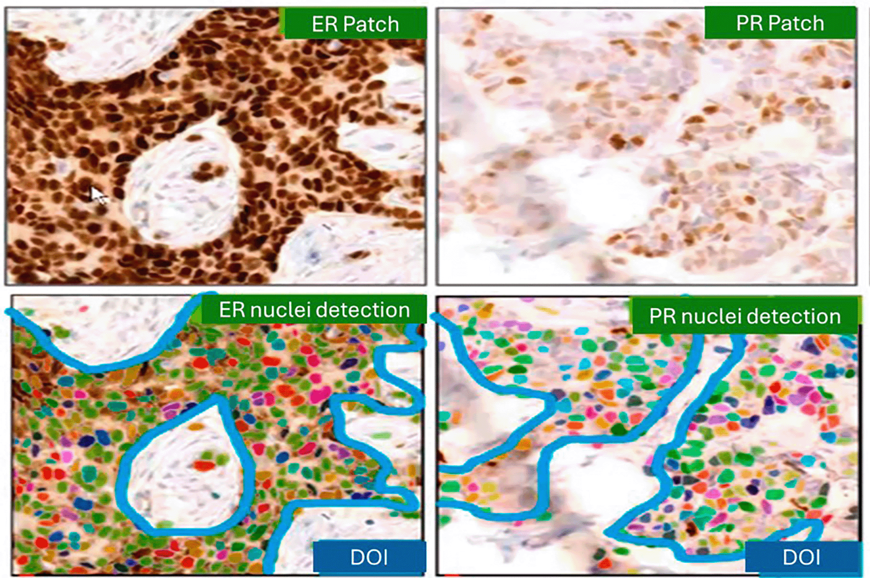
This image is output generated by the algorithm, which was developed by training AI tool (Mimansa) by pathologists and software engineers who are listed as authors to detect and represent hormone receptor staining patterns.
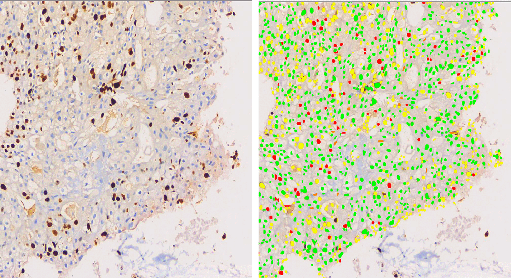
This image is output generated by the algorithm, which was developed by training AI tool (Mimansa) by pathologists and software engineers who are listed as authors to detect and represent hormone receptor staining patterns.
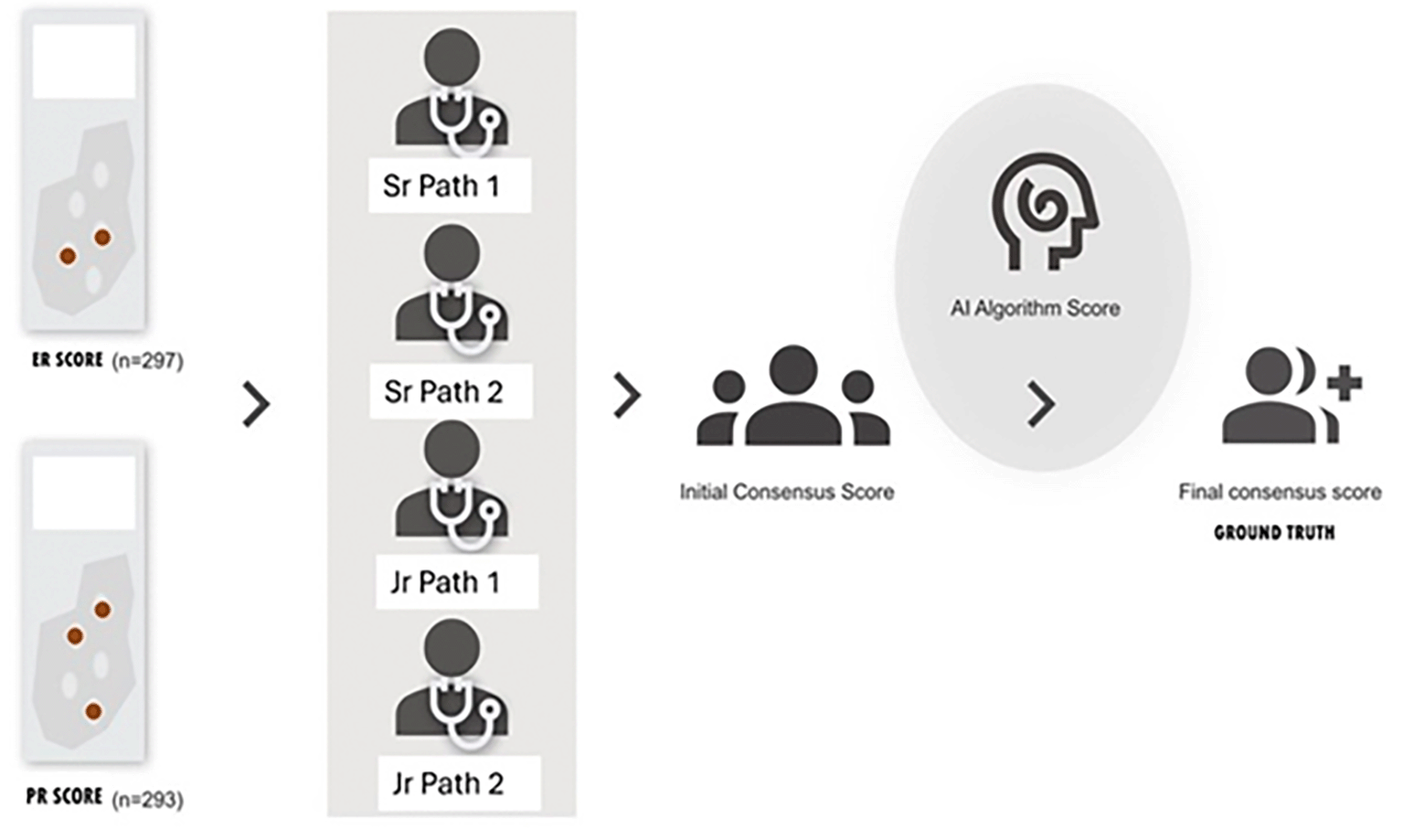
The Google material design icons were used for the figures.
Data were analyzed using IBM-SPSS Statistics for Windows version 23.0 (Armonk, NY, IBM Corp). Categorical data were expressed in terms of proportions and percentages. Inter-rater reliability analysis was performed using Cohen’s Kappa with significance testing and confidence intervals to see the agreement of the algorithm with ground truth for ER/PR scores across different score ranges as well as three groups: negative, low positive, and strong positive scores. Pearson’s correlation coefficient was used to evaluate the agreement of scores generated by the algorithm and other raters, which included senior pathologists and junior pathologists with the ground truth (final consensus scores).
The age of the patients ranged from 18 to 79 years. Trucut biopsies formed majority of cases (60.60%).
The most common histopathological diagnosis was Invasive ductal carcinoma of no special type. Luminal type showing either ER and/or PR positivity accounted tor 82.15% of the cases. Table 2 shows the pathological features of the 297 cases of breast carcinoma analysed in this study.
Table 3 reveals the performance of algorithm for ER and PR expression.and the reasons for mis-interpretation. There was concordance of 93.93% with ER expression, while slightly lower at 87.37% for PR expression scoring by algorithm when compared with ground truth. The reasons for misinterpretation are listed in Table 3 and in Figure 4.
➢ Positive staining of normal breast acini
➢ Ductal carcinoma in situ positive, while invasive tumour negative
➢ Tissue folds
➢ Stain particles
➢ Non- specific staining of stromal cells
➢ Non- specific staining of cytoplasm of tumour cells
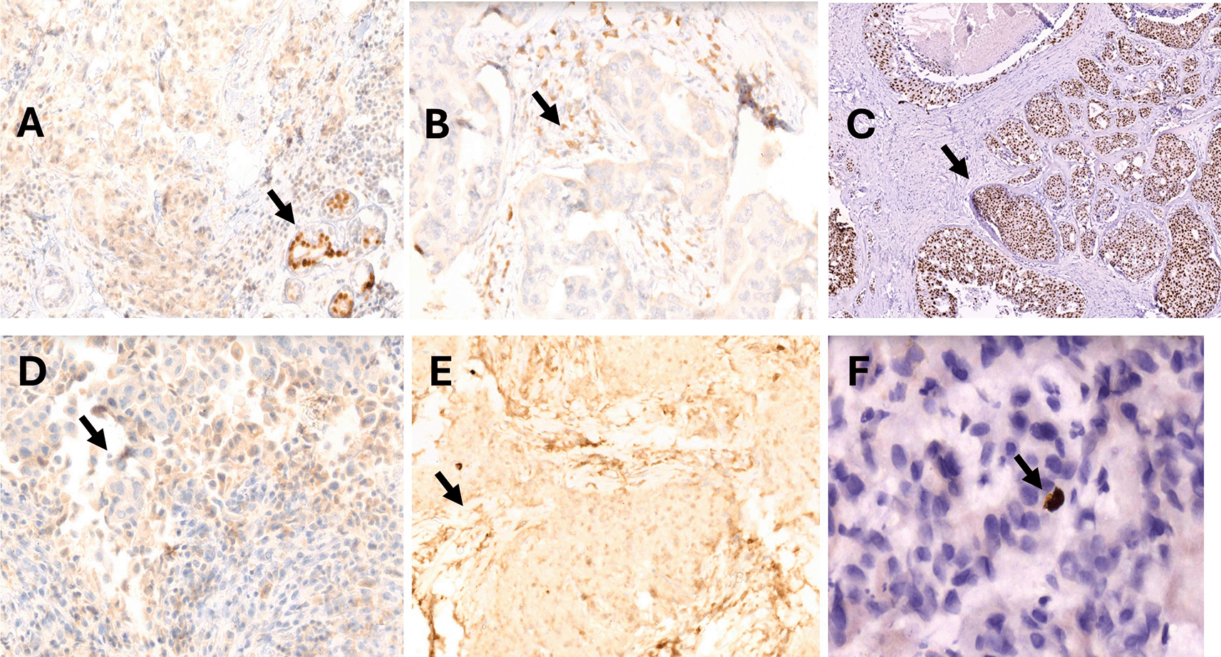
There were no statistically significant differences in ER and PR scores between the trucut biopsy and resection specimens.
Tables 4–6 reveal Algorithm performance for different scores, low positive and strong positive scores as well as comparison with senior and junior pathologists for ER expression.
For ER expression, the ground truth (GT) revealed 169 strong positive, 31 low positive, and 98 negative cases. Concordance with GT was highest among senior pathologists (99% and 98%), followed by the AI algorithm (97%), and slightly lower for junior pathologists (95% and 93%). The algorithm was particularly accurate in correctly classifying all strong positive cases; however, discordance was observed in 16 low-score cases, including four false negatives and ten false positives. Importantly, the algorithm correctly identified two true positive cases that were missed by all pathologists.
Tables 7–9 reveal Algorithm performance for different scores, low positive and strong positive scores as well as comparison with senior and junior pathologists for PR expression.
The above results for PR immunohistochemistry reveals 98% and 97% agreement for senior pathologists, 92% for the algorithm, and 93% and 91% for junior pathologists with ground truth. The algorithm for PR expression also achieved perfect concordance in strong positive cases but showed reduced performance in low-score cases, resulting in 16 false negatives and eight false positives. Cohen’s kappa coefficients demonstrated excellent agreement for ER (κ = 0.91) and strong agreement for PR (κ = 0.84).
Artificial intelligence (AI) has increasingly been applied across various domains of medical imaging, including image reconstruction, processing, analysis, and predictive modeling.10
In breast cancer, AI technologies have been widely adopted for early detection through AI-assisted radiology and for diagnostic assessment and biomarker evaluation in digital pathology.11 Appropriate image preprocessing and augmentation strategies have been shown to improve feature extraction and enhance the performance of deep learning models in medical image analysis.12
Assessing estrogen receptor (ER) and progesterone receptor (PR) status by immunohistochemistry in breast cancer is essential, as it guides treatment decisions and predicts therapeutic responses.13–15 However, the reliability of assay results depends on both the consistency of assay performance and the accuracy of its interpretation.16 Automated immune-stainers with high reproducibility, combined with whole slide digitalization and dedicated software helps in objective image quantification through color and intensity segmentation, allowing for unbiased scoring.17
Several studies have validated the performance of automated scoring methods against manual assessment of estrogen receptor (ER) and progesterone receptor (PR) expression in breast cancer and have shown a strong correlation between automated image analysis and manual scoring techniques, suggesting that automated methods can serve as reliable alternatives.17–19 Some models were based on tissue microarrays, while others utilized whole slide images for analysis.17,20,21 Some of the algorithms used required training and inputs from pathologists, while others have used algorithms that do not require supervision.22,23 Automated or digital image analysis has several advantages, such as reducing the bias in sampling, reducing inter- and intra-reader variability, providing more consistent reporting, and significantly reducing pathologists’ workload.24
In our study, we demonstrated that the algorithm developed (Mimansa) was able to detect the relevant tumor regions from WSI, quantify immunohistochemical expression, generate ER and PR scores, and effectively replicate the ER and PR scores produced by pathologist visual scoring. Our study showed good agreement of the AI algorithm in assessing ER and PR scores with the ground truth, which was slightly lower than that of pathologists. There was no statistically significant difference between the scores of senior and junior pathologists as well as the algorithm. The overall agreement between the ER score and ground truth was 93.93%, and the PR score was 87.37%. Similar results were noted in a study by Jung et al., which showed 93% concordance for ER expression (197 cases) and 89.4% (199 cases) for PR expression by the algorithm.25 Shafi et al. observed 93.85 concordance for ER expression in their study on 97 cases26 and the Pearson correlation coefficient (PCC) score in our study was 0.937 and 0.887 for ER and PR, respectively, which was similar to the study by Bankhead et al., which showed a PCC of 0.908 and 0.862 for ER and PR, respectively.27 Sharangpani et al. found an agreement of 85% and 81% between the automatic determination of positivity/negativity of ER and PR-stained cells with manual scoring.18 Gokhale et al. reported 95% concordance between automated and manual scoring, while Mofidi et al. demonstrated a highly significant correlation (r2 = 0.844) between digital and manual ones.7,17
In our study, the algorithm demonstrated excellent agreement for negative (0–2) and strong positive (6–8) groups, with kappa values of .891 and .921 for ER expression and .829 and .856, respectively, for PR expression, and moderate agreement for the low positive (3–5 scores) group with a kappa of .587 for ER expression and .534 or PR expression, suggesting the need for refinement of the algorithm to identify low positive (3–5) scores. We identified reasons for the inaccuracies in this group with false-positive scores due to misinterpretation that occurred due to interspersed positive staining of normal breast acini, ductal carcinoma in situ component when the invasive component was negative, tissue folds with brown staining, stain artifact clumps on the tissue, non-specific staining of stromal cells, and cytoplasm of the cytoplasm of tumor cells. Shafi et al. also reported three cases of false-positive ER expression by DIA, which was mainly due to intermixed benign glands in the tumor area, ductal carcinoma in situ (DCIS) components, and tissue folding.24 However, these false-positive scores in our case could be mitigated by incorporating an option in the algorithm to manually exclude areas such as tissue folds, normal breast acini, etc. before re-running the analysis. Such misclassification errors occurring from poorly stained samples or samples of bad quality can be overcome by the ability of digital image analysis to reclassify or drop individual detected objects and recalculate the software provided results.28
We noticed 7 false-negative cases for ER and 18 for PR, with the majority occurring in the low positive score range, highlighting the need for further refinement in this category. Conversely, the AI algorithm identified two true-positive ER cases that were missed by all the pathologists. This capability highlights the potential of the algorithm to significantly influence patient treatment decisions by detecting subtle positive findings that could otherwise be overlooked. This ability of the algorithm to detect critical diagnostic errors demonstrates its potential for improving diagnostic accuracy. Studies have shown improved intra- and interobserver agreement by providing pathologists with computer-aided IHC measurements during the visual scoring process. It is possible that the pathologist missed some positive cells because of the sheer size of the images.21 Since consensus scoring by experts is impractical in routine practice, automated IHC measurements may provide a means to improve scores. Shafi et al. demonstrated increased efficiency in the ER assessment of breast cancer by integrating DIA in the workflow of pathologists.26 AI analyzer could be used as an aid to pathologists as a ‘second reader’ in harmonizing judgments that may diverge due to over- or underestimations.26 Jung et al. suggested utilizing the AI analyzer as a tool for second opinions, where pathologists can maintain their original workflow, requiring reinterpretation in only a selected subset of cases (approximately 10% for ER and PR).25
Our algorithm is designed to operate on digitized whole-slide images generated through standard digital pathology workflows. Following slide scanning, the AI automatically analyzes the tumour region and generates ER and PR scores, which can then be reviewed and verified by the pathologist. The optimized two-stage architecture reduces computational time, making the workflow feasible for laboratories equipped with digital pathology infrastructure.
Limitations: One of the drawbacks of the study was the scope of validation of the AI algorithm, which was primarily confined to enhancing the concordance of pathologist interpretations. Clinical validation, such as its impact on patient survival outcomes, was not performed. Multicentre studies were included in the next version involving diverse patient populations, scanners, and staining protocols to establish the generalizability and robustness of the algorithm, however was a limitation in this study as external data sets were not used.
The version of the algorithm presented in the paper did not incorporate a tissue-type classification module to distinguish invasive carcinoma from DCIS or normal breast acini. Based on the observed misclassifications, the model is being further trained using extensive annotations to recognize and exclude ER/PR-positive normal breast acini and DCIS from analysis. Additionally, the next version will allow users to manually deselect these regions before re-running the algorithm to obtain ER and PR scores for invasive tumor alone.
The algorithm showed good agreement in scoring ER and PR expressions with the ground truth, making it a reliable tool for aiding diagnostic decision-making. It can serve as a valuable support tool for pathologists and provide a second opinion, particularly in low-positive cases where human error might occur. Notably, the algorithm identified two cases in which patients could benefit from anti-estrogen treatment, highlighting its potential clinical impact.
The study has been approved by the Kasturba Medical College and Kasturba Hospital Institutional Ethics Committee (IEC) (Ref: IEC No 05/2020 dated 18th June 2020 and extension (Amendment) IEC No 186/2021, dated 10th Feb 2021).
As the study is retrospective does not involve any intervention of subjects and uses lab based coded data collection; Consent waived by the ethics committee.
Industry Grant (APPLIED MATERIALS INDIA) Grant No: MUIND 10002256 towards development of Artificial Intelligence based decision support system for breast cancer diagnosis
The funders had no role in study design, data collection and analysis, decision to publish, or preparation of the manuscript.
© 2026 Pai K et al. This is an open access article distributed under the terms of the Creative Commons Attribution License, which permits unrestricted use, distribution, and reproduction in any medium, provided the original work is properly cited.
Current Reviewer Status: ?
Key to Reviewer Statuses VIEW HIDE
ApprovedThe paper is scientifically sound in its current form and only minor, if any, improvements are suggested
Approved with reservations A number of small changes, sometimes more significant revisions are required to address specific details and improve the papers academic merit.
Not approvedFundamental flaws in the paper seriously undermine the findings and conclusions
Version 2
VERSION 2
PUBLISHED 03 Aug 2026
Revised
Reviewer Report 06 Aug 2026
Chiman Haydar Salh, Erbil Polytechnic University, Erbil, Iraq
Approved
VIEWS 0
Competing Interests: No competing interests were disclosed.
CloseReviewer Report 05 Aug 2026
Amjad Rehman, Prince Sultan University, Riyadh, Saudi Arabia
Approved
VIEWS 0
Competing Interests: No competing interests were disclosed.
Reviewer Expertise: rMedical image analysis and cancer diagnosis
CloseVersion 1
VERSION 1
PUBLISHED 11 May 2026
Reviewer Report 14 Jul 2026
Amjad Rehman, Prince Sultan University, Riyadh, Saudi Arabia
Approved with Reservations
VIEWS 0
Is the work clearly and accurately presented and does it cite the current literature?
Yes
Is the study design appropriate and is the work technically sound?
Partly
Are sufficient details of methods and analysis provided to allow replication by others?
Partly
If applicable, is the statistical analysis and its interpretation appropriate?
Partly
Are all the source data underlying the results available to ensure full reproducibility?
Yes
Are the conclusions drawn adequately supported by the results?
Partly
References
1. Karimi M, Karimi Z, Khosravi M, Delaram Z, et al.: Feature Selection Methods in Big Medical Databases:A Comprehensive Survey. International Journal of Theoritical & Applied Computational Intelligence. 2025. 181-209 Publisher Full TextCompeting Interests: No competing interests were disclosed.
Reviewer Expertise: rMedical image analysis and cancer diagnosis
CloseReviewer Report 16 Jun 2026
Chiman Haydar Salh, Erbil Polytechnic University, Erbil, Iraq
Approved with Reservations
VIEWS 0
Is the work clearly and accurately presented and does it cite the current literature?
Partly
Is the study design appropriate and is the work technically sound?
Yes
Are sufficient details of methods and analysis provided to allow replication by others?
Yes
If applicable, is the statistical analysis and its interpretation appropriate?
Yes
Are all the source data underlying the results available to ensure full reproducibility?
Partly
Are the conclusions drawn adequately supported by the results?
Yes
Competing Interests: No competing interests were disclosed.
CloseOpen Peer Review
Alongside their report, reviewers assign a status to the article:
| Invited Reviewers | ||
|---|---|---|
| 1 | 2 | |
| Version 2 (revision) 03 Aug 26 | read | read |
| Version 1 11 May 26 | read | read |
Sign up for content alerts
Alongside their report, reviewers assign a status to the article:
Approved - the paper is scientifically sound in its current form and only minor, if any, improvements are suggested
Approved with reservations - A number of small changes, sometimes more significant revisions are required to address specific details and improve the papers academic merit.
Not approved - fundamental flaws in the paper seriously undermine the findings and conclusions